Peripheral smear call thread #3: Lymphocytosis #hemepath @pathelective
First question when approaching a smear with lymphocytosis: is it reactive or neoplastic? Patient history can provide some clues (younger patient, symptoms of infection may favor reactive).
In general, reactive lymphocytes are more polymorphous than neoplastic lymphocytes. In this patient with EBV infection, the lymphocytes vary in size and shape, and have increased amounts of pale blue cytoplasm. A rare plasma cell is also seen. 


This is a large reactive lymphocyte with immunoblast morphology. The cytoplasm is deeply basophilic and the chromatin is condensed. These can be seen in viral infections, including hantavirus and COVID-19.
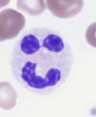
Bordetella pertussis can cause a distinctive-appearing lymphocytosis. The lymphocytes have condensed chromatin, scant cytoplasm, and clefted nuclei. Consider this diagnosis in pediatric patients with respiratory symptoms.

Lymphoproliferative disorders are more common in adult patients. In this older adult patient with chronic lymphocytic leukemia, the lymphocytes are small and monotonous, with scant cytoplasm and coursely clumped (“soccer ball” or “cracked mud”) chromatin.
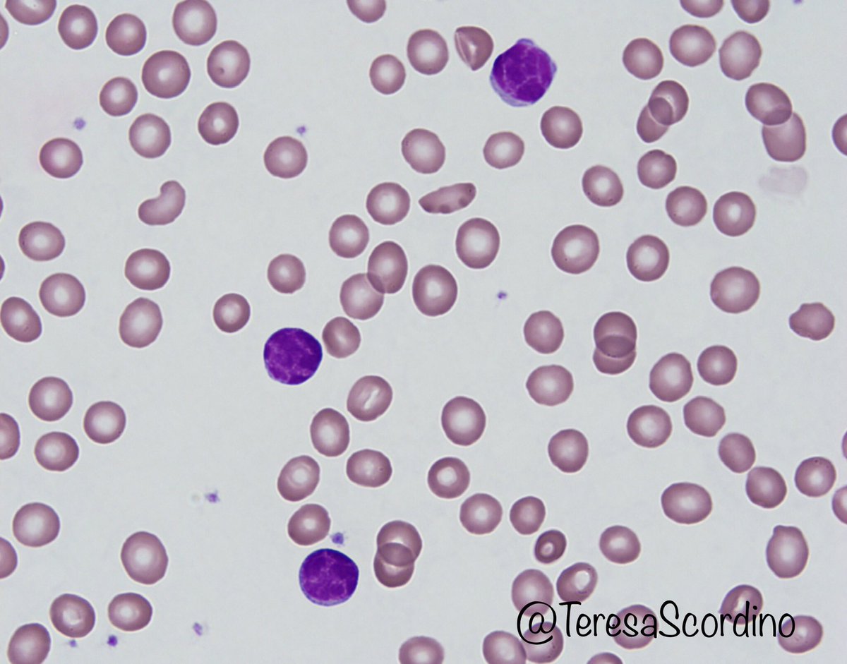
Marked lymphocytosis in a patient with B-cell prolymphocytic leukemia. The prolymphocytes have abundant cytoplasm and prominent nucleoli. The condensed chromatin helps differentiate them from blasts. Mantle cell lymphoma can also have this appearance and needs to be excluded.

The cells in T-cell prolymphocytic leukemia are small to intermediate in size, have condensed chromatin and often have visible nucleoli. Cytoplasmic blebs are frequently present. T-PLL is a mature T-cell neoplasm that is most commonly CD4+ but may also be CD4+CD8+ or rarely CD8+.

Cytoplasmic projections can be seen in hairy cell leukemia, hairy cell variant, and SMZL. Hairy cell leukemia presents with pancytopenia, but hairy cell variant often presents with lymphocytosis. HCL-v cells often have prolymphocyte-like nucleoli.


Some lymphomas (including Burkitt and mantle cell) can have a blastoid appearance. In this case of Burkitt lymphoma, the cytoplasmic vacuoles are a clue to the diagnosis (but are not entirely specific).

Remember - it’s OK to give a differential diagnosis and defer to flow cytometry if you can’t make a definitive call on morphology.