Tired of irradiating your patients with CT scans🙋♀️? Learn Renal Ultrasound #POCUS!
1⃣Perform Renal Ultrasound
2⃣Download Hydronephrosis PDF Guide!
3⃣Renal Pathology
4⃣Renal Hemodynamics (RRI/Venous Doppler)
✅New Blog Post! 👉🔗pocus101.com/Renal
#medtweetorial (1/25)👇
1⃣Perform Renal Ultrasound
2⃣Download Hydronephrosis PDF Guide!
3⃣Renal Pathology
4⃣Renal Hemodynamics (RRI/Venous Doppler)
✅New Blog Post! 👉🔗pocus101.com/Renal
#medtweetorial (1/25)👇

1 Download the FREE Hydronephrosis Grading PDF Pocket Guide! 👉🔗pocus101.com/Renal

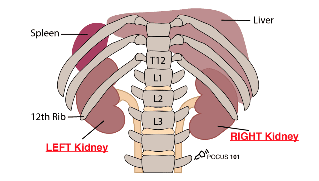
2 The kidneys are retroperitoneal organs located on either side of the vertebral column from T12-L3. Notice that the right kidney is slightly more posterior than the left kidney because of the larger size of the liver relative to the spleen. 👉🔗pocus101.com/Renal
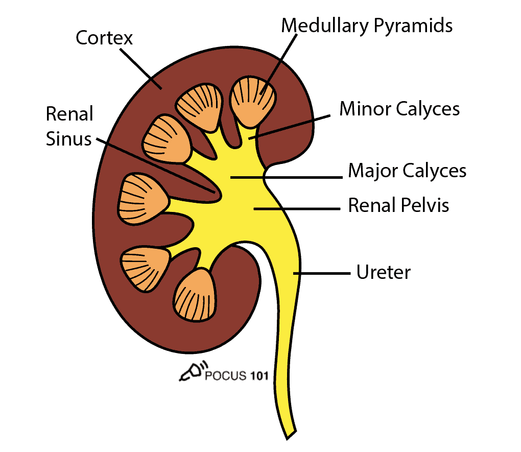
3 To perform renal ultrasound here are the key renal anatomic structures you should know:
Renal Cortex
Medullary Pyramids
Minor Calyces
Major Calyces
Renal Pelvis
Renal Sinus
👉🔗pocus101.com/Renal
Renal Cortex
Medullary Pyramids
Minor Calyces
Major Calyces
Renal Pelvis
Renal Sinus
👉🔗pocus101.com/Renal
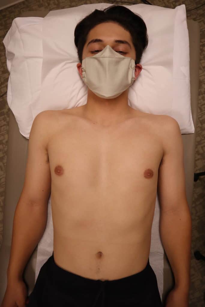
4 Right Kidney Longitudinal View:
Point probe indicator towards the patient’s head.
Place your probe at the Right Midaxillary Line around the 10th to 11th intercostal space.
👉🔗pocus101.com/Renal
Point probe indicator towards the patient’s head.
Place your probe at the Right Midaxillary Line around the 10th to 11th intercostal space.
👉🔗pocus101.com/Renal

5 Identify the: Medullary Pyramids, Renal Pelvis, Renal Sinus, Mainor Calyces, Minor Calyces, and Renal Cortex.
*The normal size of an adult kidney is around 10-11cm in the longitudinal view.
👉🔗pocus101.com/Renal
*The normal size of an adult kidney is around 10-11cm in the longitudinal view.
👉🔗pocus101.com/Renal

6 Slowly tilt/fan the probe anteriorly and posteriorly to assess the entire kidney.
👉🔗pocus101.com/Renal
👉🔗pocus101.com/Renal
7 Right Kidney Transverse View:
*Maintaining the longitudinal view of the right kidney, center the kidney on your screen, and then rotate your probe 90 degrees counterclockwise.
*The probe indicator should be pointing Posteriorly.
👉🔗pocus101.com/Renal
*Maintaining the longitudinal view of the right kidney, center the kidney on your screen, and then rotate your probe 90 degrees counterclockwise.
*The probe indicator should be pointing Posteriorly.
👉🔗pocus101.com/Renal
8 Here is the ultrasound image transition that you should see as you go from the longitudinal kidney ultrasound view to the transverse view.
👉🔗pocus101.com/Renal
👉🔗pocus101.com/Renal
9 In the transverse view, identify the Renal Cortex, Medullary Pyramid, and Renal Pelvis.
Tilt the probe superiorly and inferiorly to assess the right kidney.
👉🔗pocus101.com/Renal
Tilt the probe superiorly and inferiorly to assess the right kidney.
👉🔗pocus101.com/Renal


10 Left Kidney Longitudinal View:
*Point the probe indicator towards the patient’s head.
*Place your probe at the Left Posterior Axillary Line around the 8th to 10th intercostal space.
*Knuckles should touch the bed.
👉🔗pocus101.com/Renal
*Point the probe indicator towards the patient’s head.
*Place your probe at the Left Posterior Axillary Line around the 8th to 10th intercostal space.
*Knuckles should touch the bed.
👉🔗pocus101.com/Renal

11 Left Kidney Transverse View:
*Maintaining the longitudinal view, then rotate your probe 90 degrees counterclockwise.
*The indicator should be pointing Anteriorly.
*Tilt the probe superiorly and inferiorly to assess the entire left kidney.
👉🔗pocus101.com/Renal
*Maintaining the longitudinal view, then rotate your probe 90 degrees counterclockwise.
*The indicator should be pointing Anteriorly.
*Tilt the probe superiorly and inferiorly to assess the entire left kidney.
👉🔗pocus101.com/Renal
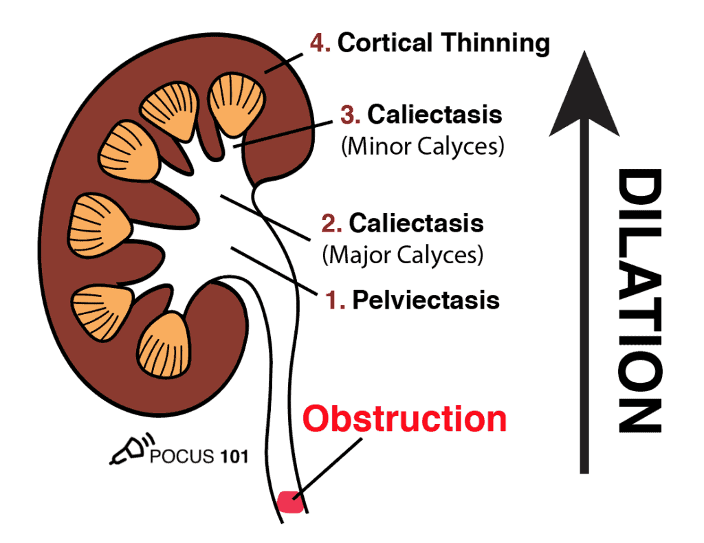
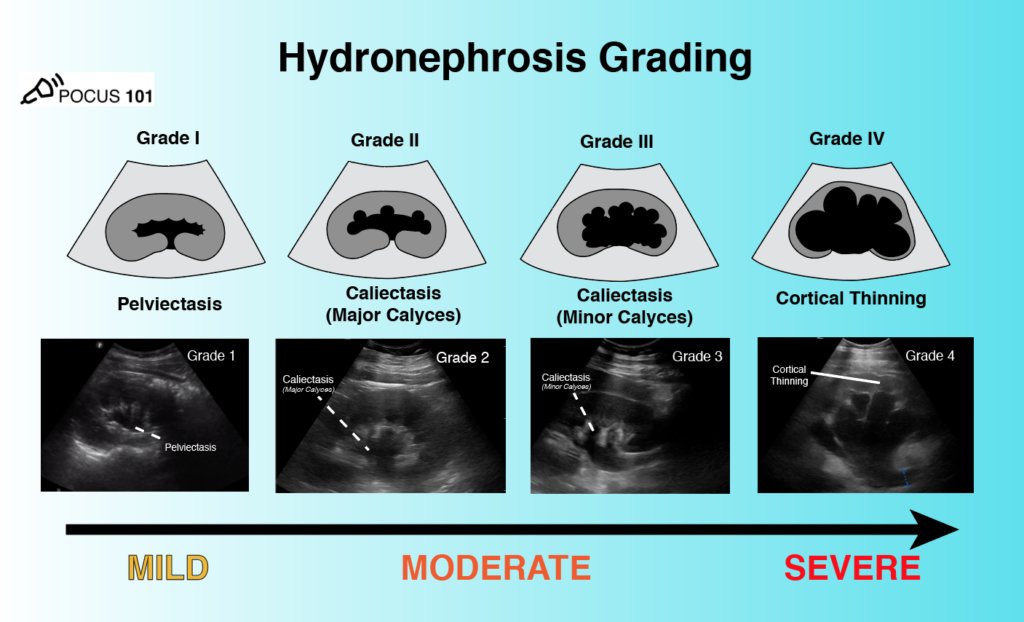
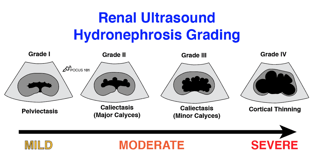
12 Don’t memorize the hydronephrosis grading system, just think that the structures closest to the obstruction will be the first to be dilated.
So the order of hydronephrosis will be Renal Pelvis -> Major Calyces -> Minor Calyces -> Renal Cortex.
👉🔗pocus101.com/Renal
So the order of hydronephrosis will be Renal Pelvis -> Major Calyces -> Minor Calyces -> Renal Cortex.
👉🔗pocus101.com/Renal

14 Size Matters!
The cutoff size for a significant kidney stone size is around 5mm. Stones <5mm have a significantly higher chance (81-98%) of spontaneously passing while stones >5mm will have a lower chance (9-65%) of passing (Jendeberg 2010).
👉🔗pocus101.com/Renal
The cutoff size for a significant kidney stone size is around 5mm. Stones <5mm have a significantly higher chance (81-98%) of spontaneously passing while stones >5mm will have a lower chance (9-65%) of passing (Jendeberg 2010).
👉🔗pocus101.com/Renal

15 Sometimes renal stones can be seen in the renal cortex, pelvis, or the ureteropelvic junction. Look for a hyperechoic structure with Acoustic Shadowing.
👉🔗pocus101.com/Renal
👉🔗pocus101.com/Renal

16 If a suspected kidney stone is lodged within the ureter or ureterovesical junction, you may find an absence of ureteral jets on the obstructed side when scanning the bladder.
👉🔗pocus101.com/Renal
👉🔗pocus101.com/Renal
17 The Twinkling Artifact is a sonographic artifact located behind calcifications of ureteral calculi when color Doppler is applied. It will appear as a multicolored high-intensity signal, similar to signals produced by turbulent flow with aliasing.
👉🔗pocus101.com/Renal
👉🔗pocus101.com/Renal


18 Renal Cyst Ultrasound Findings
*Thin-walled and smooth, without septations or any internal elements.
*The anechoic areas of cysts are localized and do not connect to the calyces.
*Round or oval in shape
*Are located in the cortex or by the pelvis.
👉🔗pocus101.com/Renal
*Thin-walled and smooth, without septations or any internal elements.
*The anechoic areas of cysts are localized and do not connect to the calyces.
*Round or oval in shape
*Are located in the cortex or by the pelvis.
👉🔗pocus101.com/Renal
19 If you see multiple renal cysts, this could indicate Polycystic Kidney Disease (PCKD) or acquired renal cystic disease (ARCD). Image from @thepocusatlas!
20 Renal Cell Carcinoma (RCC)
*Most common Renal Malignancy in Adults.
*Highly variable with regards to size, shape, and location.
*Variable echogenicity: isoechoic, hypoechoic, or echoic
👉🔗pocus101.com/Renal
*Most common Renal Malignancy in Adults.
*Highly variable with regards to size, shape, and location.
*Variable echogenicity: isoechoic, hypoechoic, or echoic
👉🔗pocus101.com/Renal

21 Angiomyolipoma
*Most common Benign Renal Tumor. (Tublin, 2011)
*Clearly circumscribed (i.e. angiomyolipoma) or irregular in shape (i.e. renal cell carcinoma).
*In some circumstances, renal cell carcinoma and angiomyolipoma can look very similar
👉🔗pocus101.com/Renal
*Most common Benign Renal Tumor. (Tublin, 2011)
*Clearly circumscribed (i.e. angiomyolipoma) or irregular in shape (i.e. renal cell carcinoma).
*In some circumstances, renal cell carcinoma and angiomyolipoma can look very similar
👉🔗pocus101.com/Renal


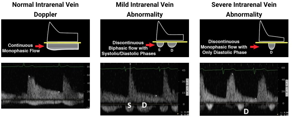
23 Below is a quick summary on how to interpret the intravenous venous Doppler tracings. #vexus
👉🔗pocus101.com/Renal
👉🔗pocus101.com/Renal
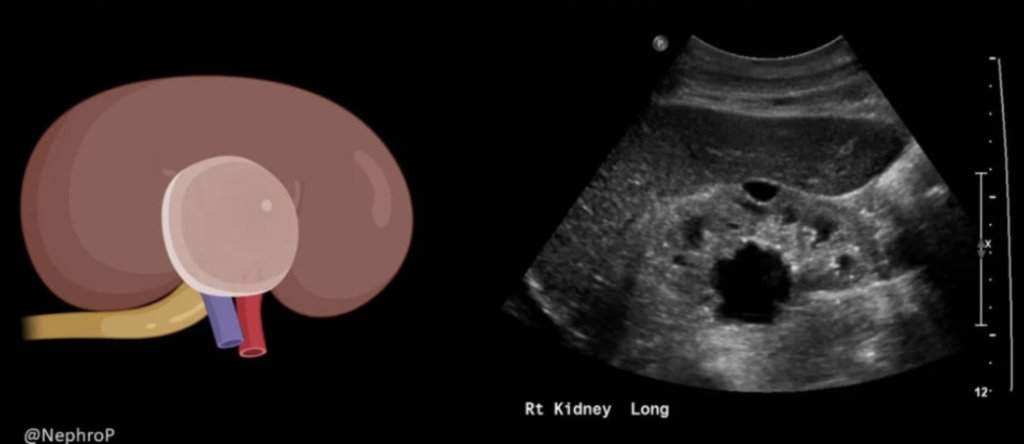
24 Recognize Hydronephrosis Mimics!
Extrarenal Pelvis
Parapelvic Cysts
Prominent Vasculature or Vascular Malformation
👉🔗pocus101.com/Renal
Figures from @NephroP, visit his website to learn even MORE about them and renal ultrasound. He is the Kidney Jedi!
Extrarenal Pelvis
Parapelvic Cysts
Prominent Vasculature or Vascular Malformation
👉🔗pocus101.com/Renal
Figures from @NephroP, visit his website to learn even MORE about them and renal ultrasound. He is the Kidney Jedi!


25 Want to learn even MORE about Renal ultrasound. Check out our Ultrasound Review book. The Renal Chapter written by @thepocusatlas @EMedCurious @MedakSD!
👉🔗pocus101.com/RevBook
👉🔗pocus101.com/RevBook

• • •
Missing some Tweet in this thread? You can try to
force a refresh