Mark Powell Profile picture
Feb 1 14 tweets 4 min read
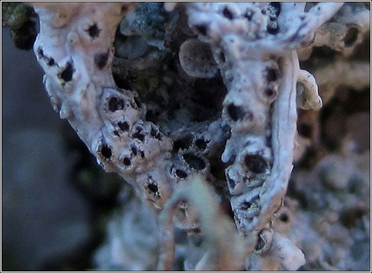
I have been sent a very interesting query from a correspondent who found this lichen on a fragment of ironstone lying on the ground. This thread will illustrate the value of recording what you observe, even if you can't name the lichen.
My correspondent assigned a provisional genus - Lecania. I think it belongs in a different genus but it is useful to make an initial hypothesis and then to keep an open mind.
Perhaps my correspondent was led astray by a misinterpretation of the spores as being septate. Spores in fresh material are difficult to interpret without 'clearing' and while iodine is a useful stain (used here), it is not harsh enough on its own to clear spores.
Perhaps these spores would appear simple if they were mounted in nitric acid, or just mounted in water and heated over e.g. a tea light candle flame.
My correspondent noticed and documented a feature displayed by some of the apothecia, which a less enquiring observer would not have considered worth noting.
Here is one of the micrographs my correspondent sent, a section through one of the apothecia, which appears to be developing a new exciple within the old apothecium.
The habitat and appearance strongly suggest the genus Trapelia to me. A C+ red reaction and presence of Chlorella type photobiont (binary fission) would help to confirm the genus.
I would never have known about the regeneration of apothecia in some species of Trapelia if it weren't for recent work by Alan Orange. He described T. collaris as new to science in 2018, naming it after the collar of old tissue surrounding the newly initiated structure inside.
The new Trapelia key uses this trait as one of the characters.
britishlichensociety.org.uk/sites/www.brit…
I can't provide a definite id. for my correspondent. The recent taxonomic work has refined the concepts but muddied the waters for field recorders and amateurs without access to molecular methods.
I haven't got my head fully around the new concepts, and regenerating apothecia are not diagnostic for T. collaris, just particularly common in that species. It is quite possible that my correspondent has found this recently described species but I can't confirm.
Despite the frustration for both of us about not having a definite name, I am always heartened when I received such well observed queries which frequently lead to important discoveries (eventually).
The reported size of the spores is well below the range for the likely Trapelia candidates. I will check with my correspondent about calibration. If they really are small, perhaps abnormally small. I don't place too much weight on spore size in most cases, using many other...
...characters in addition to simple spore size (having experience of how variable spore size can be even in a single specimen).

• • •

Missing some Tweet in this thread? You can try to force a refresh
 

Keep Current with Mark Powell

Mark Powell Profile picture

Stay in touch and get notified when new unrolls are available from this author!

Read all threads

This Thread may be Removed Anytime!

PDF

Twitter may remove this content at anytime! Save it as PDF for later use!

Try unrolling a thread yourself!

how to unroll video
  1. Follow @ThreadReaderApp to mention us!

  2. From a Twitter thread mention us with a keyword "unroll"
@threadreaderapp unroll

Practice here first or read more on our help page!

More from @obfuscans3

Feb 1
A few more parmelioids (Parmelia and related genera) from my walk today.
Can you detect the yellowish tinge in this Flavoparmelia caperata (pure grey Hypotrachyna at top right for comparison). Image
Flavoparmelia caperata in lower half, Hypotrachyna revoluta above. Again the subtle difference in colour which can only be accurately judged when the lichens are dry (they all become greener and rather similar in colour when damp/wet). Image
Here we have the two British Flavoparmelia species side by side. Similar colour. F. caperata (left) tends to be more robust, soredia coarser, produced in pustular soralia. If in doubt a simple chemical test will distinguish them. Image
Read 13 tweets
Feb 1
When dealing with mature specimens, Ramalina farinacea (left) can usually be readily distinguished from Evernia prunastri (right) and learning to separate them is a good exercise for beginners and improvers.
Thread. Image
Evernia has a slightly matt texture (perhaps like fine tissue) whereas Ramalina is more cartilaginous (as if moulded in plastic). The undersides of Evernia branches are usually starkly different (white) to the upper side. In Ramalina underside similar or only slightly paler. Image
The soralia of Ramalina farinacea are usually neatly oval in shape and restricted to edges of branches. The soralia of Evernia are irregular in shape and are not restricted to edges of branches. Image
Read 5 tweets
Feb 1
Today's gravestone, Joseph Tomlin, d. 1855, oolitic limestone. 'Only' 18 lichens plus one LF, but something a bit different, mossy and with a taxonomic conundrum.
Thread. Image
Four lichens grew upon bryophytes on this stone: Agonimia, Bilimbia, Diploschistes and Lepraria lobificans. The first two in particular are usually found on moss.
The Weddellomyces was parasitic on Caloplaca flavescens. Image
Verrucaria macrostoma f. furfuracea is a curious looking thing, the sort of lichen that would be difficult to key out. Most people get to know it by being shown it, or recognising it from photos. Image
Read 5 tweets
Jan 31
Today's gravestones, a headstone and a footstone, both limestone, Eliz. Packwood, 1917. A couple of dozen species, one of which a lichenicolous fungus.
Thread:
Plus Verrucaria polysticta on the footstone. 10/24 are members of the Verrucariaceae. C. dichroa described as new in 2006. V. obfuscans added to British list in 2015.
Three lichens, all common on the headstone, none of which would have been named correctly (or at all) twenty years ago. Caloplaca dichroa and Verrucaria ochrostoma on left side. But what about the ashy grey expanse on the right?
Read 15 tweets
Jan 31
We tend to record Diploscistes muscorum when parasitic on Cladonia (as here, from dorsetnature.co.uk/pages-lichen/l…) and D. scruposus when growing directly on rock. There are supposed to be microscopic differences but my observations seem difficult to resolve with the literature.
Thread:
Today I came across a colony growing on moss, no Cladonia in sight, on a gravestone. The number of spores per ascus fits muscorum while the septation of the spores fits scruposus! This not the first time I have noticed this problem.
The asci are consistently 4-spored which is supposed to be a D. muscorum trait.
Read 5 tweets
Jan 31
Catillaria nigroclavata is familiar to me these days on twigs and branches in my region. Will I learn any more about it if I attempt to make some drawings of it?
At first sighting, C. nigroclavata (in lower part of image) is very similar to Amandinea punctata (above). With careful study differences in appearance of both thallus and apothecia become apparent.
The thallus of A. punctata consists of minute grey warts, its apothecia become somewhat convex with maturity. C. nigroclavata has a thin, scurfy, dark, dull green thallus; the apothecia have narrow margins and discs remain flatter with age.
Read 7 tweets

Did Thread Reader help you today?

Support us! We are indie developers!


This site is made by just two indie developers on a laptop doing marketing, support and development! Read more about the story.

Become a Premium Member ($3/month or $30/year) and get exclusive features!

Become Premium

Don't want to be a Premium member but still want to support us?

Make a small donation by buying us coffee ($5) or help with server cost ($10)

Donate via Paypal

Or Donate anonymously using crypto!

Ethereum

0xfe58350B80634f60Fa6Dc149a72b4DFbc17D341E copy

Bitcoin

3ATGMxNzCUFzxpMCHL5sWSt4DVtS8UqXpi copy

Thank you for your support!

:(