1) SUMMA SUMMARUM 2.0
SPIKE PROTEIN FIBROSIS SYNDROME:
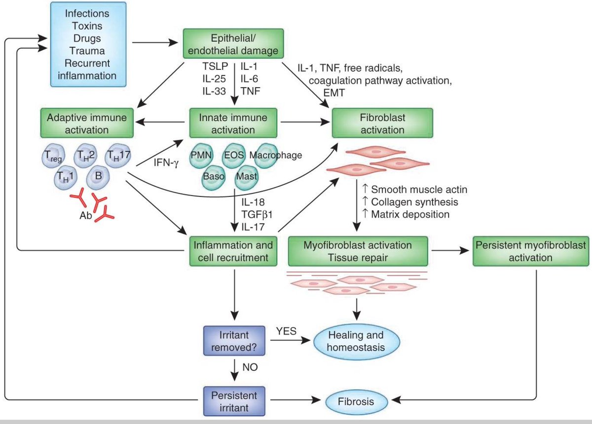
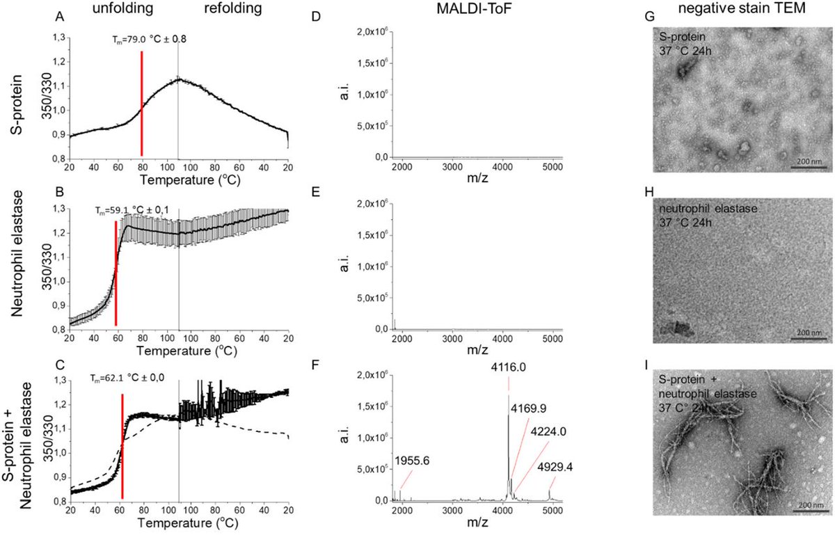
THE SPIKE PROTEIN INDUCES A FIRBROTIC CASCADE EXACTLY PARALLEL TO RADIATION FIBROSIS SYNDROME
THE SPIKE PROTEIN INDUCES THE SAME ACCUMULATION OF EXCESS FIBRIN THAT RADIATION DOES!
First: In the microcirculation, SARS-CoV-2
SPIKE PROTEIN FIBROSIS SYNDROME:
THE SPIKE PROTEIN INDUCES A FIRBROTIC CASCADE EXACTLY PARALLEL TO RADIATION FIBROSIS SYNDROME
THE SPIKE PROTEIN INDUCES THE SAME ACCUMULATION OF EXCESS FIBRIN THAT RADIATION DOES!
First: In the microcirculation, SARS-CoV-2

2) and the S protein directly enhance platelet activation and fibrin aggregation, predisposing 30–50% of COVID-19 patients to develop thrombotic events.
Various pathophysiological mechanisms have been postulated for RFS including induction of free radical (FR)-mediated DNA damage
Various pathophysiological mechanisms have been postulated for RFS including induction of free radical (FR)-mediated DNA damage
3) and subsequent apoptosis as a predisposing event.[6] Pohlers et al. described three histopathological phases of RFS such as (1) prefibrotic phase comprising ENDOTHELIAL CELLS, (2) fibrotic phase of active fibrosis containing myofibroblasts, and (3) fibroatrophic phase
4) characterized by subsequent loss of parenchymal cells. Radiation-induced (SPIKE PROTEIN) accumulation of excess fibrin in the extravascular, intravascular, and perivascular compartments has been described for RFS. Ionizing radiation (SPIKE PROTEIN) may directly result in RFS
5) by causing VASCULAR ENDOTHELIAL INJURY and indirectly by activating the inflammatory, epithelial regeneration, and tissue remodeling pathways and the coagulation cascade. Another important event is the activation of Janus kinase (JAK) and signal transducer and activator of
6) transcription (STAT) proteins along with nuclear factor kappa-light-chain-enhancer of activated B-cell (NF-KB) pathways by radiation resulting in the release of pro-inflammatory cytokines and growth factors.
NOW! Now we can unite CANCER, NEURODEGENERATION AND CARDIOVASCULAR
NOW! Now we can unite CANCER, NEURODEGENERATION AND CARDIOVASCULAR
7) DISEASE that has heretofore appeared as RANDOM. But, it is nothing of the sort!
CANCER
This Spike Protein Fibrosis Syndrome not only explains the cancer we are seeing, but also its AGGRESSIVENESS. Tumors are characterized by extracellular matrix (ECM) deposition, remodeling,
CANCER
This Spike Protein Fibrosis Syndrome not only explains the cancer we are seeing, but also its AGGRESSIVENESS. Tumors are characterized by extracellular matrix (ECM) deposition, remodeling,
8) and cross-linking that drive fibrosis to stiffen the stroma and promote malignancy. The stiffened stroma enhances tumor cell growth, survival and migration and drives a mesenchymal transition. A stiff ECM also induces angiogenesis, hypoxia and compromises anti-tumor immunity.
9) Not surprisingly, tumor aggression and poor patient prognosis correlate with degree of tissue fibrosis and level of stromal stiffness. In this review, we discuss the reciprocal interplay between tumor cells, cancer associated fibroblasts (CAF), immune cells and ECM stiffness
10) in malignant transformation and cancer aggression.
NEURODEGENERATION
The process of uncontrolled internal scarring, called fibrosis, is now emerging as a pathological feature shared by both peripheral and central nervous system diseases. In the CNS, damaged neurons are not
NEURODEGENERATION
The process of uncontrolled internal scarring, called fibrosis, is now emerging as a pathological feature shared by both peripheral and central nervous system diseases. In the CNS, damaged neurons are not
11) replaced by tissue regeneration, and scar-forming cells such as ENDOTHELIAL CELLS, inflammatory immune cells, stromal fibroblasts, and astrocytes can persist chronically in brain and spinal cord lesions. Although this process was extensively described in acute CNS damages,
12) novel evidence indicates the involvement of a fibrotic reaction in chronic CNS injuries as those occurring during neurodegenerative diseases, where inflammation and fibrosis fuel degeneration.
Of course, the cardiovascular implications of fibrosis are widely established and
Of course, the cardiovascular implications of fibrosis are widely established and
13) do not need to be recapped here.
I believe we are dealing with a progressive fibrotic syndrome that starts in the microvasculature. This also explains all of Long COVID.
Clearly, all Spike Protein accelerants must be stopped IMMEDIATELY.
ncbi.nlm.nih.gov/labs/pmc/artic…
I believe we are dealing with a progressive fibrotic syndrome that starts in the microvasculature. This also explains all of Long COVID.
Clearly, all Spike Protein accelerants must be stopped IMMEDIATELY.
ncbi.nlm.nih.gov/labs/pmc/artic…
14) onlinelibrary.wiley.com/doi/full/10.10…
pubmed.ncbi.nlm.nih.gov/34671772/
frontiersin.org/articles/10.33…
ncbi.nlm.nih.gov/labs/pmc/artic…
pubmed.ncbi.nlm.nih.gov/34671772/
frontiersin.org/articles/10.33…
ncbi.nlm.nih.gov/labs/pmc/artic…
• • •
Missing some Tweet in this thread? You can try to
force a refresh


