1) BUILDING: MY MOST IMPORTANT FINDING TO DATE: DIED UNEXPECTEDLY SOLVED
SUDEP AND “DIED UNEXPECTEDLY” – THE SPIKE PROTEIN, FIBROSIS AND THE BRAINSTEM
PART I “A SILENT ENTRANCE”
As you recall, the original spike caused a loss of the sense of smell, the medical term is Anosmia.


SUDEP AND “DIED UNEXPECTEDLY” – THE SPIKE PROTEIN, FIBROSIS AND THE BRAINSTEM
PART I “A SILENT ENTRANCE”
As you recall, the original spike caused a loss of the sense of smell, the medical term is Anosmia.


2) The olfactory (sense of smell) system happens to be a DIRECT ROUTE TO THE BRAINSTEM.
Indeed, when we look at autopsies, the Spike Protein is a very frequent “guest” in the Brainstem. Pathological immune responses or SARS-CoV-2 invasion of the brainstem is suspected.
Indeed, when we look at autopsies, the Spike Protein is a very frequent “guest” in the Brainstem. Pathological immune responses or SARS-CoV-2 invasion of the brainstem is suspected.
3) An autopsy study has isolated 32 brain sections from 16 victims of COVID-19 and found concentrated SARS-CoV-2 RNA (>5 copies/mm3) in three sections from the olfactory nerves and the brainstem’s MEDULLA. More convincingly, in another autopsy study of deceased COVID-19 patients,
4) SARS-CoV-2 RNA and proteins (nucleocapsid or SPIKE) were detected in 50% and 40% of brainstem samples, respectively. Similarly, another autopsy study has found SARS-CoV-2 RNA and SPIKE PROTEINS in the olfactory mucosal-neuronal junction and brainstem’s MEDULLA in 67% and 19%
5) of samples, respectively. In sum, these autopsy studies have provided evidence for SARS-CoV-2 tropism FROM THE OLFACTORY SYSTEM INTO THE BRAINSTEM.
OK. We know the Spike Protein invades the Medulla of the Brainstem. So, what does this mean? Well, those who suffer from Epilepsy
OK. We know the Spike Protein invades the Medulla of the Brainstem. So, what does this mean? Well, those who suffer from Epilepsy
6) are prone to sudden death. In fact, it is because of PATHOLOGIC CORRELATIONS IN THE MEDULLA that they suffer what is known as SUDEP- Sudden Unexpected Death in Epilepsy.
Sudden unexpected death in epilepsy (SUDEP) likely arises as a result of AUTONOMIC DYSFUNCTION around the
Sudden unexpected death in epilepsy (SUDEP) likely arises as a result of AUTONOMIC DYSFUNCTION around the
7) time of a seizure. In vivo MRI studies report volume reduction in the MEDULLA and other brainstem autonomic regions. Rostro-caudal alterations of medullary volume in SUDEP localize with regions containing respiratory regulatory nuclei. They may represent seizure-related
8) alterations, relevant to the pathophysiology of SUDEP.
PART II “FIBROSIS AND SUDEP”
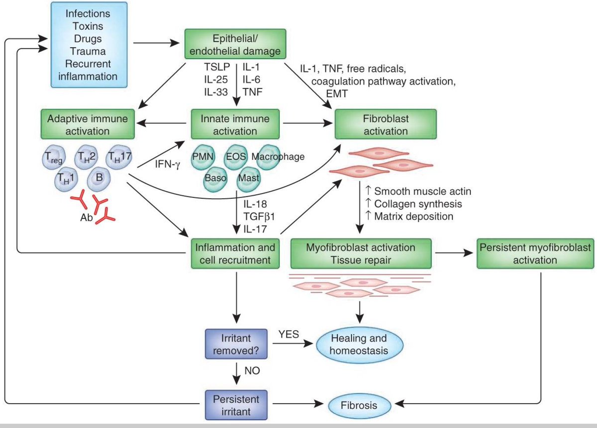
Now, this heretofore rare event is now becoming all too common. The “One-Two Punch” of aberrant spike protein brainstem signaling (remodeling) combined with spike protein cardiac remodeling
PART II “FIBROSIS AND SUDEP”
Now, this heretofore rare event is now becoming all too common. The “One-Two Punch” of aberrant spike protein brainstem signaling (remodeling) combined with spike protein cardiac remodeling
9) has virtually recreated the EXACT ENVIRONMENT OF SUDEP!
The histologic evaluation was possible in 65% of the cases (15/23) whose death was attributed to SUDEP and in 71% (15/21) of controls. Forty percent of the SUDEP cases (6/15) presented several foci of fibrotic changes in
The histologic evaluation was possible in 65% of the cases (15/23) whose death was attributed to SUDEP and in 71% (15/21) of controls. Forty percent of the SUDEP cases (6/15) presented several foci of fibrotic changes in
10) the deep and subendocardial myocardium in contrast to 1 control (6.6%, P = 0.03). None of the subjects from the SUDEP group showed fibrotic changes in their conduction system as compared with 1 control (6.6%). The quantitative evaluation of fibrosis demonstrated a trend
11) toward more fibrosis in the deep and subendocardial myocardium of the SUDEP cases. Forty percent of cases in the SUDEP group were men (6/15), characteristically young at time of death (mean age 38 years) and with a late epilepsy onset (mean age 21 years). Antemortem, 73% of
12) the SUDEP patients (11/15) had experienced infrequent seizures (self-reported). We conclude that the SUDEP cases displayed significant fibrosis of the myocardium when this was assessed by qualitative means. This fibrosis may be the consequence of myocardial ischemia as a
13) direct result of repetitive epileptic seizures, which, associated with the ictal sympathetic storm, may lead to lethal arrhythmias.
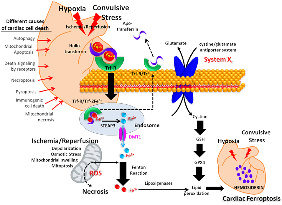
And frontiersin.org/articles/10.33…, the Spike Protein’s induction of Iron Overload plays into this.
And frontiersin.org/articles/10.33…, the Spike Protein’s induction of Iron Overload plays into this.
14) I implore all public health authorities: The Spike Protein Accelerants must be stopped this instant.
researchgate.net/publication/22…
pubmed.ncbi.nlm.nih.gov/32559314/
pubmed.ncbi.nlm.nih.gov/15897710/
frontiersin.org/articles/10.33…
https://t.co/gx1CEQcgcs
researchgate.net/publication/22…
pubmed.ncbi.nlm.nih.gov/32559314/
pubmed.ncbi.nlm.nih.gov/15897710/
frontiersin.org/articles/10.33…
https://t.co/gx1CEQcgcs
• • •
Missing some Tweet in this thread? You can try to
force a refresh


