
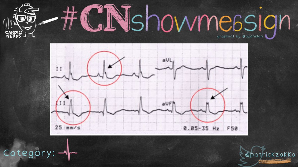
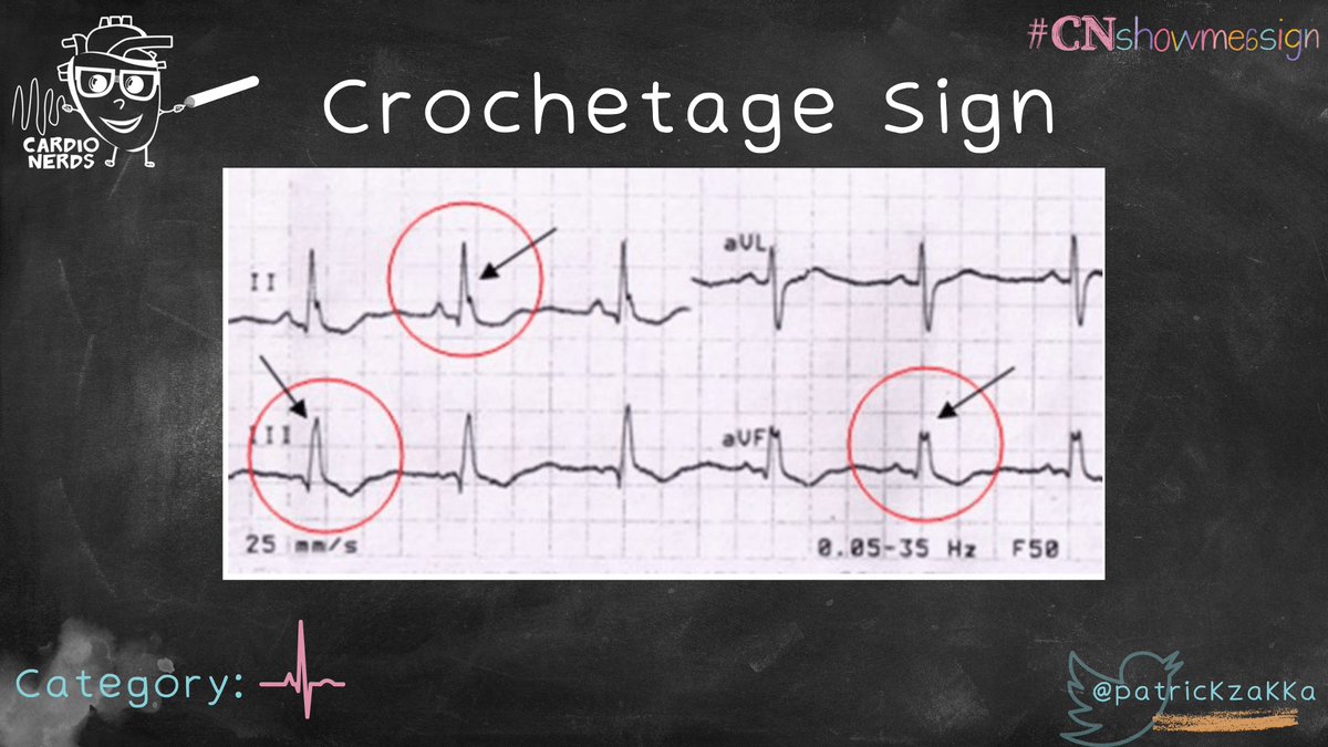
Crochetage sign is a notch on the apex of R waves in the inferior leads. It can be associated with ostium secundum ASDs. 
It is a result of delayed depolarization of the right ventricle because of L to R shunt. This is why ostium secundum ASDs are also associated with:
🚦Right axis deviation
🚦Incomplete RBBB
🚦Right axis deviation
🚦Incomplete RBBB
⚡️ Sens/spec: 73.1%/92.6% (when in 1 inferior lead); 27.8%/100% (when in all inferior leads). Increased specificity when associated with incomplete RBBB.
⚡️ Correlated with greater anatomic defect or greater L to R shunt.
⚡️ Sens/spec for dx PFO in cryptogenic stroke: 36%/91%.
⚡️ Correlated with greater anatomic defect or greater L to R shunt.
⚡️ Sens/spec for dx PFO in cryptogenic stroke: 36%/91%.
⏳ "Crochetage" = French for "notching"
⌛️ First described 1959 by Rodriguez-Alvarez et al in 11 patients with ostium secundum ASDs.
⌛️ First described 1959 by Rodriguez-Alvarez et al in 11 patients with ostium secundum ASDs.
References:
pubmed.ncbi.nlm.nih.gov/8613618/
casereports.bmj.com/content/2016/b…
Thank you to my mentor @jcurrier17 for teaching me about EKGs seen in ASDs and inspiring this med ed project.
pubmed.ncbi.nlm.nih.gov/8613618/
casereports.bmj.com/content/2016/b…
Thank you to my mentor @jcurrier17 for teaching me about EKGs seen in ASDs and inspiring this med ed project.
#CNshowmeasign brought to you by @PatrickZakka and @a_h_ghoneem.
Special thank you to @TDonisan (for the amazing graphics) and @AmitGoyalMD @Dr_DanMD for reviewing.
If there are any other signs you would like us to tackle, put them in the comments and let us know what to do next!
Special thank you to @TDonisan (for the amazing graphics) and @AmitGoyalMD @Dr_DanMD for reviewing.
If there are any other signs you would like us to tackle, put them in the comments and let us know what to do next!

• • •
Missing some Tweet in this thread? You can try to
force a refresh


