1) A short 🧵looking at some key points for Lumbar Spinal Stenosis (LSS)
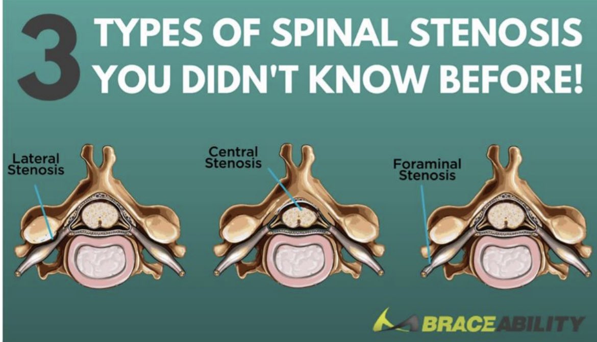
2) LSS is defined as narrowing of the spinal canal +/- neural foramina which causes crowding around the exiting caudal nerve roots with compression & ischaemia due to progressive, age related changes to the anatomical structures of the spine.
3) Epidemiology: approx 103 million people suffer with symptomatic LSS with 20% of > 60yrs suffering with it, yet 80% are thought to be asymptomatic (Katz et al., 2022). Other research found a higher prevalence and ? 45-60% of adults >65yrs (Comer et al., 2020).
4) Pathophysiology; DDD puts greater load on posterior facet joints which alongside the ligaments hypertrophy and this encroaches on surrounding nerves. Pain is thought to be from ischaemia to small arterioles of the nerve +/- venous congestion. Most common is L4/5 (Lee et al 20)
5) Features; UL/BL pain in the LL (rarely the feet). Sx relief w flexion or walking flexed. LBP, neuro Sx when walking or standing for long period & normal symmetrical dorsalis pedis pulses (Genevay et al., 2018). Chronic Sx can impede balance & mobility.
6) Diagnosis: Can be made through clinical assessment. Previously Cluster of Cook was found to have a high LR if pt’s had 4/5 (Cook et al 2011). More recently N-Class Criteria (see pictures) has been proposed but there is little research on its psychometric properties.
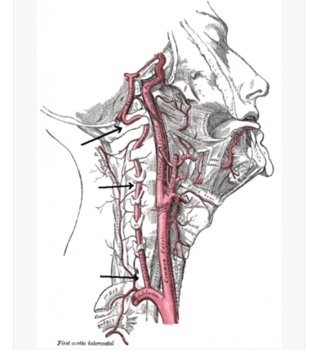
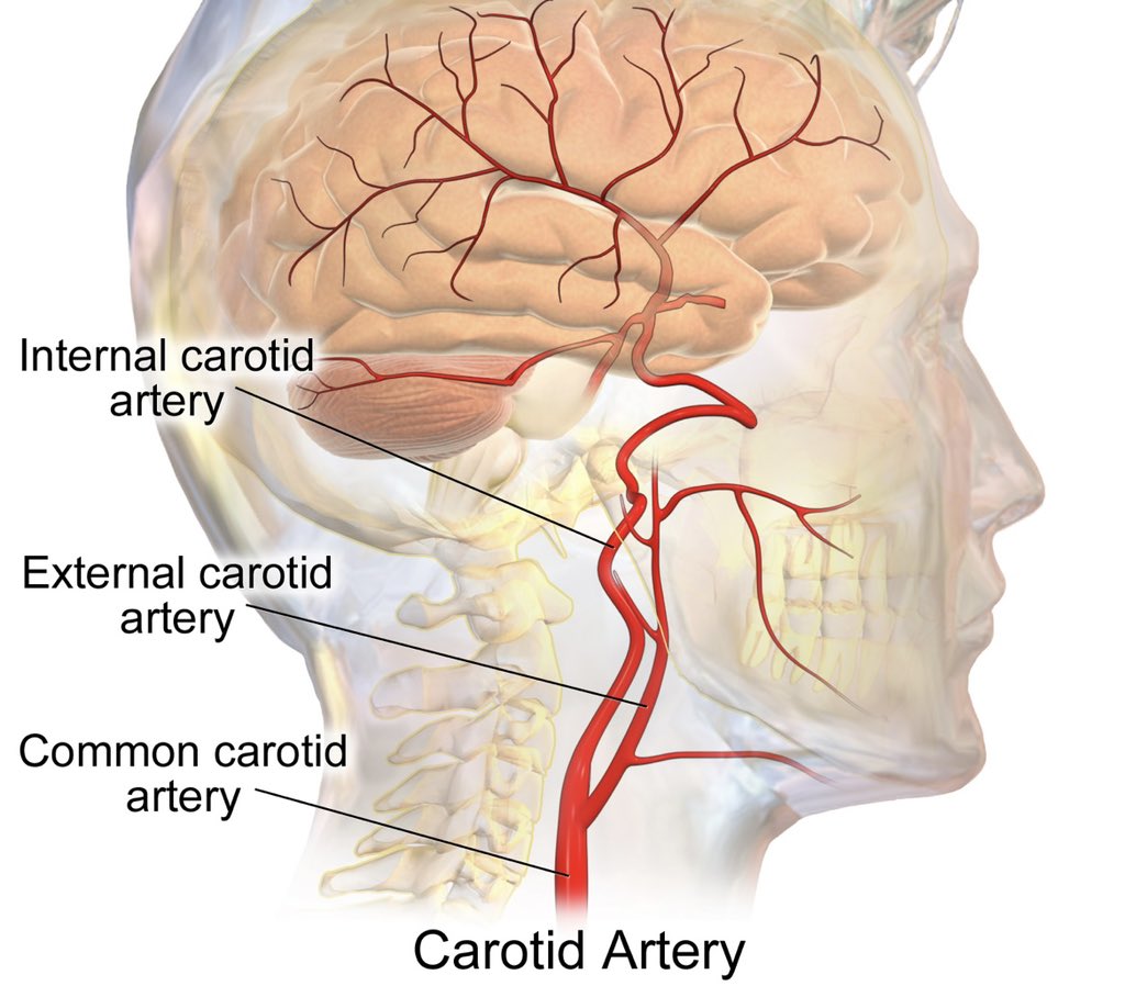
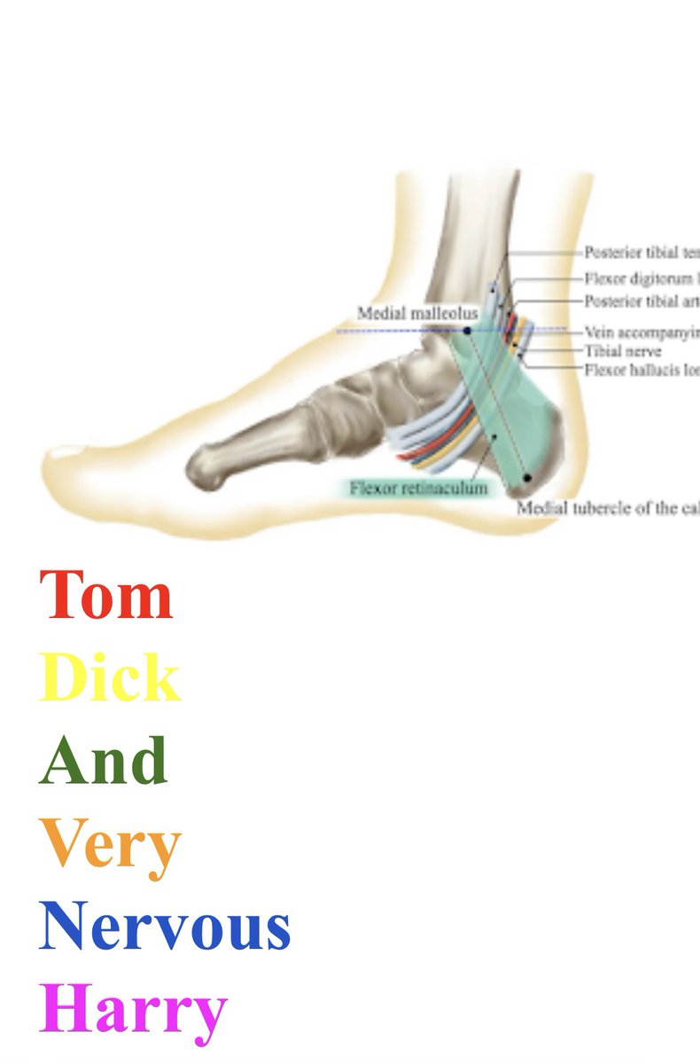
7) PAD - a differential Dx which can elicit similar Sx. Clinicians should be aware of vascular comorbidities, smoking Hx & physically assess for LL oedema / skin integrity, pulses & Buerglers. Sx will be provoked irrespective of spinal position w PAD’s during exertion (Bike Test)
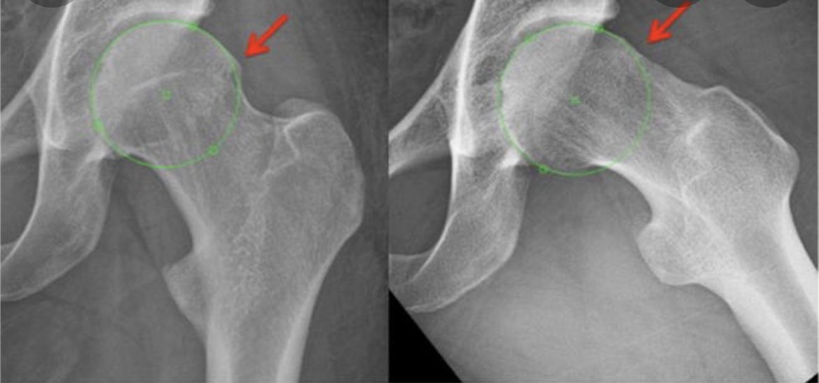
8) Imaging - low correlation between Sx & findings. Utilise for pt’s with persistent Sx that are impeding function. MRI can be used to assess the diameter of the spinal canal. CT w myelogram can provide more accurate info w dynamic positions (flex vs ext) - Lee et al., 2020.
9) Prognosis: A study of 146 pt’s found a 1/3 improved, 50% remained ISQ & 10-20% became worse with symptoms at 3yr f/u (Wessberg et al., 2017).

researchgate.net/publication/31…
10) Treatment; Host of different techniques. There has been some low evidence supporting the use of cycling, aquatic & supported treadmill walking. This is thought to increase the CSA of the canal and reduces axial loading on the spine. For further info this paper is great.
11) Surgery; Should only be considered when significant disability and no improvement with Rx. 10-24% can suffer complications post-decompression (Temporiti et al 2022). Those with concomitant scoliosis / spondylolithesis may require fusion to prevent spinal instability.

• • •

Missing some Tweet in this thread? You can try to force a refresh
 

Keep Current with Charlie Clements

Charlie Clements Profile picture

Stay in touch and get notified when new unrolls are available from this author!

Read all threads

This Thread may be Removed Anytime!

PDF

Twitter may remove this content at anytime! Save it as PDF for later use!

Try unrolling a thread yourself!

how to unroll video
  1. Follow @ThreadReaderApp to mention us!

  2. From a Twitter thread mention us with a keyword "unroll"
@threadreaderapp unroll

Practice here first or read more on our help page!

More from @ClementsCharl96

May 2
1) A 🧵 providing a whistle stop tour of Frozen Shoulder (FS)
2) FS is associated with stiffness of the capsule which restricts GHJ A/PROM within a capsular pattern coupled with a normal XR finding (Zuckerman 2011). Previously thought to be self-limiting, now ++ evidence highlighting the significance it has on pain, disability and function
3) Epidemiology: Thought to affect 2-5% of the gen pop between the age of 40-70yrs. Women are more susceptible & it is thought that 6-17% of patients are unfortunate enough to develop it in the contralateral arm within the first 5yrs (Dyer et al., 2021).
Read 12 tweets
Apr 21
1) A 🧵 looking at Quadrilateral Space Syndrome (QSS). A rarer cause of shoulder pain
2) QSS is a rare condition which causes vague Sx around the shoulder (often posteriorly) that occurs secondary to compression of the axillary nerve (AN) +\- posterior humeral circumflex artery (PHCA) as they travel through this region.
3) The space is an opening in the posterior wall of the axilla. It is bordered by the teres minor (superior), teres major (inferior) the humeral shaft (lateral) & long head triceps (medial). Albeit rare, should be a differential in younger, overhead athletes
Read 11 tweets
Apr 19
1) A thread looking at Adductor Related Groin Pain (ARGP) Image
2) ARGP is the most common culprit for groin pain in athletes & can be the result of an acute strain of the myotendinous junction. Or, more persistent & associated with adductor tenderness around the insertion of the pubic bone (Holmich, 1997). Image
3) ARGP accounts for 5-18% of all sports injuries (Serner et al., 2015). It is common in kicking & multidirectional sports & accounts for 16-18% of football injuries w approx 1-1.1 per 1000hrs & a higher prevalence in men vs women (Bisciotti et al 2021; Weir et al., 2015).
Read 10 tweets
Apr 13
1) A 🧵 looking at Cervical Artery Dissection (CAD). A vascular pathology which is a differential for cervical pain that requires urgent consideration Image
2) A dissection of either the carotid/vertebral arteries. The carotid arteries are positioned around the front of the neck whilst the vertebral are posterior. Their incidence is low (2.6 per 100k) but it is the most common cause of ischaemic stroke in <50’s (Debette & Leys 2009).
3) A haematoma forms within the arterial wall (most common internal carotid), this can expand towards the intima & cause luminal narrowing/stenosis, endothelial injury & thrombus formation. Whereas bleeding which spreads towards the adventitia may weaken the artery = aneurysm. Image
Read 12 tweets
Apr 8
1) A 🧵 looking at Tarsal Tunnel Syndrome (TTS)
2) TTS is an Entrapment neuropathy of the tibial nerve & it’s branches as it passes within the tunnel, posterior & inferior to the flexor retinaculum. It’s a rare condition & doesn’t appear to be any epidemiological studies looking at its prevalence (Antoniadis & Scheglmann 2008)
3) Structures that sit within the TT are; tibialis posterior, FDL, posterior tibial artery & vein, tibial nerve and the FHL. A useful pneumonic for this is - ‘Tom, Dick And Very Nervous Harry’.
Read 13 tweets
Apr 4
1) A 🧵 overviewing Femoroacetabular Impingement of the Hip Image
2) FAI is a movement related disorder involving symptomatic contact between the femoral head & acetabulum (Griffin et al 2016). It occurs due to changes in the morphology of the hip. *Note - v common (15-25%) in asymptomatic pt’s (Dijkstra et al 21).
3) CAM Morphology - 3:1 prevalence in males (Kuhn et al 2015). Osteophytosis of the femoral head/neck due. These can occur primary (physiological response to loading) OR secondary (pre-existing hip pathology). XR for A-angle, >50-55 is considered CAM. Image
Read 12 tweets

Did Thread Reader help you today?

Support us! We are indie developers!


This site is made by just two indie developers on a laptop doing marketing, support and development! Read more about the story.

Become a Premium Member ($3/month or $30/year) and get exclusive features!

Become Premium

Don't want to be a Premium member but still want to support us?

Make a small donation by buying us coffee ($5) or help with server cost ($10)

Donate via Paypal

Or Donate anonymously using crypto!

Ethereum

0xfe58350B80634f60Fa6Dc149a72b4DFbc17D341E copy

Bitcoin

3ATGMxNzCUFzxpMCHL5sWSt4DVtS8UqXpi copy

Thank you for your support!

Follow Us on Twitter!

:(