You can see the 4 cannulae in this X-ray. The RVAD drainage is from the RA and returns to the PA. The LVAD drains LV apex and returns to ascending aorta. You’ll also notice the lungs look ok and the patient isn’t intubated….. so why the oxygenator in the RVAD…….?
This patient has a dilated cardiomyopathy and deteriorated despite inotropes so needed to be rescued with VA ECMO. Notice the stylet is pulled back during the insertion of the venous drainage cannula, there are markings on the stylet to enable this…. Did you know??
The LVEDP was measured at 40 so we also inserted a #impella and the patient made stabilised with #ecpella therapy. He went into theatre for his bivad well resuscitated and with good end organ function. Despite this he had ICU acquired weekness and so was not able to be extubated
Adding the oxygenator to the RVAD enabled us to remove CO2 and so effectively decrease his work of breathing to a level he could manage. This meant he was able to avoid a tracheotomy and so eat, drink and talk immediately. The oxygenator enabled us to effectively #humanise him.
Here is how a bivad levitronix looks without an oxygenator. Here you can see 2 blue and 2 red pipes, this time with the pumps in shot. The cannulation is identical. Notice how the LVAD chatters with the cardiac cycle.
28yo 👨🦰with end stage DCM on the urgent list for heart transplantation ( milrinone dependent, INTERMACS 3) .
VT storm today --> deterioration to INTERMACS 1 (crash and burn)
pVA ECMO inserted. Flow 4lpm
CXR post ECMO insertion
24h later.
You've already guessed what's going to happen:
🟠AV not opening, flat art line
🔴LA + LV + A root filled with smoke
🟢Severe MR
🟡 PCWP 35 ( I know, the PAC is "radiolucent" in this CXR 😅)
🔵 High VT burden, inotropes ⏫⏫⏫
🟣 ⏫ O2 requirements
What can you do?
Please answer what you would do in your center (regardless of what you think I am going to say)
Take into account this patient has end stage DCM (he is not going to come off ECMO easily)
Hi!
Ready for a VV ECMO case? 🍿
A 🧵...
54yo male,end stage IPF active on the lung transplant list.
Cannulated and retrieved from his local hospital for respiratory failure.
Post admission CXR below. What's missing in this film?
Yes, there is no ETT.
He was cannulated awake and we'll try to keep him self ventilating until the transplant surgery.
VV ECMO as a bridge to transplant is fascinating, but not easy.
There must an MDT agreement before proceeding. It doesn't work for all patients!
What next?
Day 7. paO2 is 🔽. You start troubleshooting:
ECMO/oxygenator checks:✅
ECMO blood flow increased: ✅5.5lpm, DLP= -75
Recirculation ruled out:✅
Chest imaging: ✅
Morning everyone!Curious to see what happened to our patient?
Well, 48h have gone by and she's responded very well to so far. FiO2 is 0.35, fluid balance is 5L negative and her PP is 25 mm Hg.
Let's do a sedation hold.... oh, no..
EMERGENCY BUZZER. 🧵
She is on VT with no pulsatility and MAP is 24 mm Hg.
ECMO flow 🔽 1.5 lpm and is fluctuating, drainage pressure is -160. Thomas, The ECMO CNS looking after the patient, tells you he cannot increase the flow.
A cardiac arrest call is put out
Your colleague Marina🔼 FiO2 to 100%, and assesses A + B from standard ALS, Thomas draws a blood sample for an ABG and inspects all iv infusion lines, SHO Nicki starts CPR and you are asked to look at the ECMO machine and find out what is going on. 😰
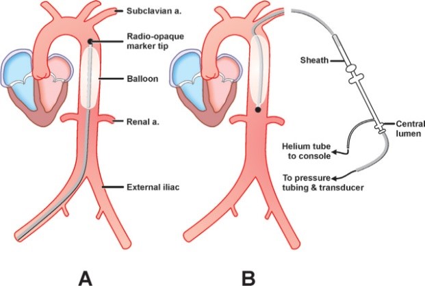
Device of the day - Intra-Aortic Balloon Pump (IABP)
The most widely used circulatory support device – cheap, low cost, with relatively few complications. The IABP is designed to be situated in the descending aorta, distal to the left subclavian artery and proximal to the...
renal arteries. It should inflate at the onset of diastole (aortic valve closure) and deflate prior to the beginning of systole.
It is used to support critically ill patients in a few ways, fundamentally providing circulatory, coronary and ventricular support...
1. Augmentation of intra-aortic pressure in diastole --> increase coronary, cerebral and systemic blood flow and perfusion 2. Presystolic IABP deflation --> decreased afterload and LVEDP. This mechanism is particularly important in acute ischaemic cardiogenic shock, where...
1️⃣ Sutures! A minimum of three sutures, well secured and appropriately positioned. (Picture is quite outdated but it shows the sutures)
2️⃣ Dressing! A transparent, chlorhexidine impregnated dressing that will allow visual inspection and measurement from insertion site to end of metal coil.
3️⃣ Tubing holder -tight enough the keep tubing safe but without causing extra pressure and allowing mobilisation (above knee)
Cardiogenic shock team assemble - a powerful coalition of specialties and disciplines able to rapidly assess and deploy the right treatment for the right patient at the right time in the right place. The patient is arriving in 10minutes……
Severe right ventricular failure on #echofirst and SCAI E+ cardiogenic shock. Placed immediately on peripheral VA ECMO