1) A short 🧵 looking at median nerve palsy (MNP) and some useful tips to help differentiate the site of its lesion.
2) In terms of its anatomy. It is composed of two sections, the lateral cord (C5-7) which innervates proximal muscles and provides sensory innervation to thenar eminence and the radial 3.5 digits. The medial cord (C8-T1) is purely motor.
3) 1st if we start with the carpal tunnel (CT) which accounts for most MNP’s. An easy way to determine whether the lesion is proximal, is if the pt is c/o paraesthesia in the thenar eminence as this is innervated by the palmar cutaneous branch found 2 inches proximal to wrist.
4) Useful clues to help determine CT vs C5/6 radiculopathy would be; a) +’ve flick sign, +’ve phalens & tinels, often nocturnal paraesthesia and wrist flexion + triceps would be normal!
5) If there has been a lesion proximal to the elbow (e.g supracondylar #) we will lose all MN innervation. Easiest way to spot this is that the forearm will be supinated with an inability to pronate due to denervation of pronator teres & quadratus.
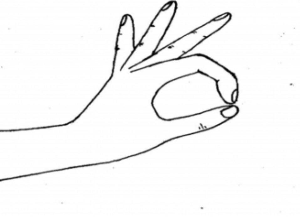
6) If we have an anterior interosseous lesion (albeit rare) this will only affect the medial cord and therefore we are presented with motor deficits. Inability / weakness with pincer grip / performing an OK sign is an easy way to assess FPL and IF flexors.
7) Another sign of AIN is the Hand of Benediction. We will see natural flexion of our ulnar sided digits. Whereas denervation of FPL, FDS and FDP will mean the thumb, IF & MF rest in an extended position.
8) Finally, with more chronic issues, denervation of the APB will mean the hand appears more flattened as the thumb will naturally lie in a more adducted position.
• • •
Missing some Tweet in this thread? You can try to
force a refresh
1) Intermittent claudication is something often encountered in practice, especially in our older demographic of patients. But how can we differentiate between neurogenic vs vascular? Or if the two occur concomitantly⁉️
2) Neurogenic claudication (NC) is associated with pain, paraesthesia, fatigue & weakness in LL & develops due to neurovascular compression, secondary to central canal stenosis. It occurs due to a reduction in CSA around the spinal canal which is exacerbated in upright positions
3) Vascular claudication (VC) is a metabolic problem whereby atherosclerotic changes to arteries restricts blood flow and causes weakness, cramping and aching in the legs during exertion.
1) Some slides I’ve put together illustrating the outcomes of surgery for LSS and considerations to make as clinicians when deciding how to optimally manage this demographic of patients
2) No hard or fast rule but these are some criteria which helps guide decision-making. (Note the top point is deemed urgent!)
1) Short 🧵 looking at the outcomes between surgery vs conservative treatment for LSS.
2) This Cochrane review consisting of 5 trials (shock low quality evidence) concluded that there was very little confidence to determine which was better & clinicians should be cautious when informing patients on potential treatment option.
3) 3 studies compared laminectomy vs conservative Rx & found no SD in ODI scores at 6/12 or 1yr follow-up. Only 1 low quality study favoured surgery at 2 years. It was difficult to draw conclusions between the results as the details of conservative Rx weren’t well described
1) A 🧵 looking at EPL ruptures following distal radius fractures (DRF).
2) DRF’s are the 2nd most common # after the hip & is the most within the upper limb. This is often a Colles which occurs secondary to a FOOSH with the forearm supinated, resulting in dorsal displacement of the radius (Corsino et al., 2022).
3) EPL rupture is fortunately rare, accounting for <1% of complications post-DRF. They appear to frequently occur around 4-8/52 after the fracture & are more common in older patients as opposed to paeds (Song et al., 2013).
1) A 🧵 looking at lumbar radicular pain & radiculopathy
2) These symptoms develop secondary to compression / irritation of the lumbosacral nerve roots. Common culprits of mechanical pressure are disc herniation (most common - often degenerative), stenosis, spondylosis. Less common can be space occupying lesions / spondylolithesis.
3) Radicular pain is classified (in simple terms) as a gain in function & is associated with ectopic signals released from the dorsal root ganglion of the nerve root. Pt’s will reporting lancinating, shooting, burning Sx due to inflammation of the affected nerve.