The Invissible 🧵Theory
Working theory, hit "Show replies" to see more
Could these white rubbery #CalimariClots found by embalmers be amyl0id fibrin clots being formed in the vasculature?
Working theory, hit "Show replies" to see more
Could these white rubbery #CalimariClots found by embalmers be amyl0id fibrin clots being formed in the vasculature?
https://twitter.com/ScotteSwz/status/1549477928446169089
What if the COVID vaxx (V) mRNA doesn't always stay in the muscle but travels in the🩸where it transfects the endothelial cells in the vasculature and organs?
"Here we provide evidence that circulating SARS-CoV-2 sp!ke proteins are present in the plasma🩸of participants
"Here we provide evidence that circulating SARS-CoV-2 sp!ke proteins are present in the plasma🩸of participants
vaxxinated with the mRNA V...
These data show that S1 antigen production after the initial V can be detected by day 1 and IS PRESENT BEYOND THE SITE OF INJECTION AND THE ASSOCIATED REGIONAL LYMPH NODES." academic.oup.com/cid/article/74…
These data show that S1 antigen production after the initial V can be detected by day 1 and IS PRESENT BEYOND THE SITE OF INJECTION AND THE ASSOCIATED REGIONAL LYMPH NODES." academic.oup.com/cid/article/74…
The cells might express the sp!ke protein, a foreign protein to the immune system.
The immune cells might signal the complement system, to attack the endothelial cells, and activate the platelets.
The👇 descriptions: "rubbery" "tough" "calamari"
The immune cells might signal the complement system, to attack the endothelial cells, and activate the platelets.
The👇 descriptions: "rubbery" "tough" "calamari"
https://twitter.com/crankycrab1171/status/1539006441688039424?s=20&t=ONT3EaBQ0pvlTm1tbrSMQw
This would create a feed-forward loop of: signaling, attack, scarring, signaling, wound repair and fibrosis.
The endothelial lining would be destroyed and its cells would be soughed off.
The cells that form the inner part of the vasculature,
The endothelial lining would be destroyed and its cells would be soughed off.
The cells that form the inner part of the vasculature,
the Tunic Media are rubbery muscle cells held together with collagen and elastin to keep them tight and firm and regulate🩸pressure. 

When these cells are stripped off the vessels would become leaky, causing a further activation of the immune system.
A coagulation cascade or InflammoThromboticResponse (see work of Dr. Richard Fleming) comenses, and immune cells rush to the site to repair the vessels.
A coagulation cascade or InflammoThromboticResponse (see work of Dr. Richard Fleming) comenses, and immune cells rush to the site to repair the vessels.
Quickly amyl0id fibrin clots are produced to repair the leaks.
This is not isolated to one area, as sp!ke protein is being produced in many endothelial cells at once in all the vessels.
The signaling, attack, damage and repair cascades are going on simultaneously all along the
This is not isolated to one area, as sp!ke protein is being produced in many endothelial cells at once in all the vessels.
The signaling, attack, damage and repair cascades are going on simultaneously all along the
vessels.
This leads to a traffic jam in the vessels.
They become crowded with hyperactive accumulation of complement and coagulation cells.
Cell debris from this process accumulates and is caught up in the amyl0id fibrin cl0ts being formed.
Eventually the fibrin nets catch many
This leads to a traffic jam in the vessels.
They become crowded with hyperactive accumulation of complement and coagulation cells.
Cell debris from this process accumulates and is caught up in the amyl0id fibrin cl0ts being formed.
Eventually the fibrin nets catch many
cells and form long fibrin strands the length and shape of the veins and arteries.
🩸 would continue to flow around the flexible cl0ts which float in the vessels. Around them the capillaries are destroyed. Eventually this impeded blood flow and vascular destruction would cause
🩸 would continue to flow around the flexible cl0ts which float in the vessels. Around them the capillaries are destroyed. Eventually this impeded blood flow and vascular destruction would cause
POTS, hypoxia, tissue necrosis and thrombosis.
This is the possible cause of the amyl0id fibrin micro cl0ts found in the blood of Long Covid patients. #TeamClots is helping remove them with apheresis.
This is the possible cause of the amyl0id fibrin micro cl0ts found in the blood of Long Covid patients. #TeamClots is helping remove them with apheresis.
The fibrin cl0ts appear yellow in the aphaeresis treatment and white when extracted by embalmers


A good description of the clotting cascade of Acute Covid/Long Covid. Ddimer can only detect the initial part of this phase, eventually the clots are too large and resistant to fibrinolysis, as #TeamClots research shows.
https://twitter.com/8Oquz7wXRmQ5aaT/status/1567337703133401088?s=20&t=n2G2H2ci1Eim9_EBcIKD-g
Thioflavin T a fluorescent dye marker, is good at attaching to the micro clots as they continue forming.
https://twitter.com/doctorasadkhan/status/1485868228492640256?s=20&t=n2G2H2ci1Eim9_EBcIKD-g
#CovidIsVascular
https://twitter.com/doctorasadkhan/status/1568216217252421632?s=20&t=FEKzCdQhKNV8LCtBhqGllQ
More images of the fibrin clots formed in the blood after V
https://twitter.com/chrislittlewoo8/status/1577950371502252037?s=20&t=FEKzCdQhKNV8LCtBhqGllQ
The V clots might be different and denser than those formed in convalescent LC.
Here are some possible reasons for it and for the odd shapes.
Add to this the fact that collagen is being stripped from the tunica media. Collagen is known to form crystals.
Here are some possible reasons for it and for the odd shapes.
Add to this the fact that collagen is being stripped from the tunica media. Collagen is known to form crystals.
https://twitter.com/overcatbe/status/1575776981584592896?s=20&t=nEZsSm1ZB-mDQ5EgTkInyg
A detailed description of the sepsis ~ clot formation feed forward loop.
Spike signaling, complement destruction, bleeding, leaking, scarring, signaling, coagulation cascade:
The Spartacus Letter by @NameIsSpartacus iceni.substack.com/p/the-spartacu…
Spike signaling, complement destruction, bleeding, leaking, scarring, signaling, coagulation cascade:
The Spartacus Letter by @NameIsSpartacus iceni.substack.com/p/the-spartacu…
Some highlights:
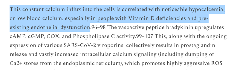
The sp!ke protein (SP) is the key that turns the lock of the cell membrane channels open in the vessels and causes destruction and flooding.
The sp!ke protein (SP) is the key that turns the lock of the cell membrane channels open in the vessels and causes destruction and flooding.

Calcium regulates every cell membrane in the body.
The SP can cause systemic destruction. This is why COVID and the COVID V have such heterogeneous symptoms.
The SP can cause systemic destruction. This is why COVID and the COVID V have such heterogeneous symptoms.

It is particularly important with regard to brain cells and amyl0idosis
pubmed.ncbi.nlm.nih.gov/35281104
pubmed.ncbi.nlm.nih.gov/35281104
Immune cells rush in (some are caught up in the amyl0id clots). Red Blood Cells are also transfected by SP and lose their 02. This is a very important point (and possibly why the #calimariclots are white?) The destruction of the RBCs means tissue necrosis is accelerated.

Necrosis already initiated by the SP's membrane permeabilization, and Ca+ flooding in the cells of surrounding tissues, but their necrosis is increased by the lack of 02.
(Interesting paper showing damaged RBCs are recycled in the liver):
sciencedaily.com/releases/2016/…
(Interesting paper showing damaged RBCs are recycled in the liver):
sciencedaily.com/releases/2016/…
4:50 Jessica Rose points out that sp!ke can bind to the RBCs and destroy the hemoglobin molecule. This causes amyl0id plaques to form.
Amyl0ids are extremely hard, dense clots. This is the form of the micro clots shown by #teamclots.
rumble.com/v1jyhpb-dr.-je…
Amyl0ids are extremely hard, dense clots. This is the form of the micro clots shown by #teamclots.
rumble.com/v1jyhpb-dr.-je…
They are what make the long fibrils in the vessels tough and resistant to fibrinolysis.
This echoes the latest research on amyl0idosis in Alzheimer's disease.
It reverses 100 years of theory, and shows that amyl0id clots form when the soluble form of amyl0id beta DECREASES.
This echoes the latest research on amyl0idosis in Alzheimer's disease.
It reverses 100 years of theory, and shows that amyl0id clots form when the soluble form of amyl0id beta DECREASES.
It's not the hard amyl0id beta plaques causing AD,
it's the decrease of the soluble form of the protein that causes disease.
The plaques are just the residue, the signature.
it's the decrease of the soluble form of the protein that causes disease.
The plaques are just the residue, the signature.

The amyl0id clots and long fibrils are formed by the destruction of the RBC's AND the sloughing off of the endothelial tunica media AND the responding immune cells AND other passing molecules in the🩸.
#calimariclots are the kitchen sink of clots.
#calimariclots are the kitchen sink of clots.
They form quickly because the SP, in a t0xin-like manner, efficiently destroys every cell it transfects.
This debris clumps together, higgledy-pigledy and forms long fibrils snaking through the vessels.
Sp!ke snake moves through the vasculature and sheds its skin as it goes.
This debris clumps together, higgledy-pigledy and forms long fibrils snaking through the vessels.
Sp!ke snake moves through the vasculature and sheds its skin as it goes.
#TheInvissibleThreadTheory #TheInvissibleThread #Ouroboros #VoodooInMyBlood #Turn2Stone #Hyperactive #SP #TheFallOut #IronMan #Amyl0idosis #MicroCl0ts #Fibrosis #CytokineStormyWeather #CD147 #Alzheimers #AlzheimersDisease #pandemicplaylist
The AD study authors make some pertinent points.
.
"I think this is probably the best proof that reducing the level of the soluble form of the protein can be T0XIC."
.
"I think this is probably the best proof that reducing the level of the soluble form of the protein can be T0XIC."

Dr. Spikelove
https://twitter.com/W777Jonathan/status/1584322887158112256?s=20&t=G0Llb3x-0bXf3ccSoEuWKg

He's been saying this for over a year and a half.
Thank you Dr Hoffe. #ToldYouSo
Thank you Dr Hoffe. #ToldYouSo
https://twitter.com/_Janey_J/status/1585352567412117504?s=20&t=SUoGRBZDtEygnMO8u14TMg
#Amyl0idosis #Amyloidfibrils #MicroClots #MedTwitter
https://twitter.com/Sunheatgenes72/status/1582143822200700928?s=20&t=SUoGRBZDtEygnMO8u14TMg
Another description of the clots:
https://twitter.com/jeffgilchrist/status/1586043146042978304?s=20&t=zjxPpTB5DPRy-mCkLQ5wWg
Along with the Thioflavin T test there is another test to detect micro clots, VQ SPECT:
https://twitter.com/drclairetaylor/status/1566178384429948928?s=20&t=zjxPpTB5DPRy-mCkLQ5wWg
#malopomalo #clotbyclot
https://twitter.com/scrowder/status/1587808355908820993?s=20&t=uqYLeQIz3TbXw8_zZjQKyA
"these could actually be fragments of elastic fibers that have somehow detached themselves from the damaged vessels' intima"
Genau Doktor Burkhardt! odysee.com/@hipsterious:3…
Genau Doktor Burkhardt! odysee.com/@hipsterious:3…
And I knew yes I knew I should run
But then I heard her say 💋 #CalimariClots
But then I heard her say 💋 #CalimariClots
https://twitter.com/ssayssayssay/status/1595169592418004992?s=20&t=MTAy0Z2j4BcmsmW_tgUmjg
"Bolus Theory" has a different explanation:
https://twitter.com/GirardotMarc/status/1595180394105692163?s=20&t=MTAy0Z2j4BcmsmW_tgUmjg
I agree that LNPs are destructive too.
https://twitter.com/GirardotMarc/status/1595181043606339584?s=20&t=MTAy0Z2j4BcmsmW_tgUmjg
Come a little bit closer doc
amidwesterndoctor.substack.com/p/what-is-caus…
amidwesterndoctor.substack.com/p/what-is-caus…
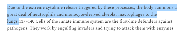
The point of the pathophyisiological mechanism of the #InvissibleThread is, it is happening to several cells in the endothelium, everywhere, AT THE SAME TIME.
The overwhelmed and hyperactive immune feed forward loop results in fibrin clots.
The overwhelmed and hyperactive immune feed forward loop results in fibrin clots.
https://twitter.com/TheoNNN22/status/1596972856436719616?s=20&t=UnD7kReXGiggehtvU_K__w
Dr. Duncan is a cardiologist w/ degrees in cardiovascular and infectious diseases. Since the pandemic began she has started Long Covid clinics and researches Long Covid.
Her description of the pathophysiology of LC amyloid fibrin micro clots is The Invissible Thread Theory. >
Her description of the pathophysiology of LC amyloid fibrin micro clots is The Invissible Thread Theory. >
"They are not normal clots...if you make too many of these clots...these clots are getting sequestered, or stuck, in the capillary vascular beds, and when you have enough of these IT IS OBSTRUCTING THE FLOW OF O2 TO EVERY SINGLE ORGAN IN THE BODY"
@ 2:40
@ 2:40
I agree w/ Dr. Duncan, what COVID/LC pathophysiology indicates is long term vascular and cardiac disability and death. Myocarditis is silent, it is also the tip of the vascular iceberg with the spike protein.
This is why for the last 2 years one of my main tags is: #3to5.
This is why for the last 2 years one of my main tags is: #3to5.


Histology slides showing contents of the clots @ 52:15
"More pale blue area down below, that's the glycoproteins, the thicker pink bands that's the fibrin and the amyloid..up above parts of it that are speckled with blue...are normal blood elements trapped within these."
"More pale blue area down below, that's the glycoproteins, the thicker pink bands that's the fibrin and the amyloid..up above parts of it that are speckled with blue...are normal blood elements trapped within these."

I believe, eventually Dr. Cole and Dr. Burkhardt may come to the same conclusion I have. #CalimariClots are being formed from the immune system's destruction of the endothelium, after its cells express the spike protein. #MedEd #MedTwitter >
https://twitter.com/HowardSteen4/status/1599517591004856320?s=20&t=2qC2ilaLRFxhHR8EbRwRFw
This reaction is exaggerated in the vaxxed because the mRNA with pseudouridine is designed as a gene therapy to be durable in the body. Thus there is more spike protein expressed, and for longer time than occurs in viral infection, creating a feed forward loop of destruction. >
Jessica Rose does a great job of breaking down the Stephanie Nye et al. paper and explaining the problem of COVID vaxx mRNA durability, codon optimization and aberrant protein expression.


So many variables with each dose, each administration of the dose and each recipient of the dose:
https://twitter.com/CanningPharm/status/1599879075073359872?s=20&t=InQIBcvsAtQ7ZKyXpCbEyg
In Part 2 of this essay Pierre Kory analyzes some of the same issues with mRNA durability that Jessica Rose does
(These are long research threads. When you reach the end hit "Show replies" to see all)
(These are long research threads. When you reach the end hit "Show replies" to see all)
https://twitter.com/PierreKory/status/1599552091067850752?s=20&t=L-ZtLEHUi8Mb7XoVqeX0DQ

This is an interesting anecdote. I wonder how many other HCWs are seeing this phenomenon in patients?
As Arne Burkardt showed, he was able to extract a #CalimariClot from the blood of the vaxxed former marathon runner who was experiencing circulation problems in her feet.
As Arne Burkardt showed, he was able to extract a #CalimariClot from the blood of the vaxxed former marathon runner who was experiencing circulation problems in her feet.

Tin in the clots:
https://twitter.com/Greg21143362/status/1599210450163757057?s=20&t=L-ZtLEHUi8Mb7XoVqeX0DQ
AD is one of the risk factors for severe COVID, and death. Do these patients have higher amounts of Tin in their bodies, which is speeding up clotting?
https://twitter.com/Greg21143362/status/1599565624124256256?s=20&t=L-ZtLEHUi8Mb7XoVqeX0DQ
Here is a "debunk" of the #CalimariClots.
Except it seems to confirm the nature of the clots.😉
Except it seems to confirm the nature of the clots.😉

This is an interesting point to consider on the lack of some elements in the clots.

"Everyone who has taken the jabs is bloodclotting...
They're cutting full blood clots out of people right now, as we speak."
He said this over a year ago.
#DDimer #Troponin #ThioflavinT
They're cutting full blood clots out of people right now, as we speak."
He said this over a year ago.
#DDimer #Troponin #ThioflavinT
https://twitter.com/liz_churchill7/status/1600202464979931136?s=20&t=L-ZtLEHUi8Mb7XoVqeX0DQ

Double Eureka
https://twitter.com/bH2Omiamigo/status/1600448882025975809?s=20&t=DMFPl2ZWeqRIeAdYB-gc9g


These two threads☝️👇 mesh.
https://twitter.com/PlanZip/status/1595272059151683587?s=20&t=DMFPl2ZWeqRIeAdYB-gc9g
Interesting question and replies.
https://twitter.com/lora2241/status/1600429034197770241?s=20&t=Pi2FQodJHLSBjmdmLO1JiA
"also frequently, after rare codon substitution, the increased yield of the recombinant protein in E. coli is accompanied by a reduction of its solubility, and its accumulation in inclusion bodies" >


This would explain the clots in a COVID convalescent person but how could it do so in a COVID vaxx person? Could the mRNA transfect bacteria in the body and do something similar?
Can this reaction happen with other microbes in the body? What about yeast. Do people who have an overabundance of these microbes in their bodies have a higher risk of amyloidosis when the spike protein (virus or vaxx) is introduced and affects protein translation?
What is clear is that the translation process in microbes is dynamic. Changes happening in real time cause new states:

A wonderful detailed @HighWireTalk with @drcole12 showing the pathology of the #CalimariClots. Supplements the research of @resiapretorius & @dbkell of #TeamClots on the amyloid fibrin nature of the clots. Evidence! #WeveGotTheSlides! Woot! 🥳
1:14:58
rumble.com/v1zn8m0-episod…
1:14:58
rumble.com/v1zn8m0-episod…
Interesting read:
https://twitter.com/DrDan20000/status/1603051675119325184?s=20&t=wEhlPGFOwCRQBQ_urQGm_Q
This premier research scientist on Alzheimer's Disease showed that the deceased AD patients all had high levels of aluminum in their brains.
https://twitter.com/HighWireTalk/status/1597992747293904901?s=20&t=Ur-CID2wyXHbQCM8m7JFgw
This seems to support our theory that metals help the amyloid protein formation.
Amy loi di Amy loi dah clot goes on bra
La la how the clot goes on
La la how the clot goes on
https://twitter.com/PierreKory/status/1612950684168589314?s=20&t=xiuy_8f4OmZqxHWiv7Sf2w

Synthetic mRNA vs viral mRNA also plays a large part in the increased amyloidosis and amyloid micro clots seen in the vaxxed, as Kevin McKernan's research shows.
open.substack.com/pub/christiela…
open.substack.com/pub/christiela…

As Pretorius and Kell's research shows, people with Diabetes already have an inflamed endothelium and micro clots. The spike protein increases this. Are these patients at risk of faster or increased thrombosis?
https://twitter.com/Ashmedaidemon/status/1617228587454853120?s=20&t=TTdtUSh5oeKbqdAzgteLUA
Long 🧵of 🧵s scroll 👆 keep showing replies
The amyloid fibrin #CalimariClots seen by embalmers, are also seen by doctors.
MDs are attempting to clear clots surgically but cannot and are amputating limbs.
🧵 of many Go Fund Me cases with the same clots:
The amyloid fibrin #CalimariClots seen by embalmers, are also seen by doctors.
MDs are attempting to clear clots surgically but cannot and are amputating limbs.
🧵 of many Go Fund Me cases with the same clots:
https://x.com/janiesaysyay/status/1701607201872511404?s=20
A 🧵 of people with multiple recurring 🩸cl0ts:
https://x.com/janiesaysyay/status/1685851100510072833?s=20
• • •
Missing some Tweet in this thread? You can try to
force a refresh