Cerclage wires got a good wrap in 2022. Several conference presentations, a couple of posters and abstracts, and now here's a mini literature collection for @DrMarecek and myself to bath in the next time the topic comes up #orthotwitter
just an aside, i was honestly a bit skeptical whether people would follow links to off-site content, and looking at the 30 odd likes on this tweet i was thinking "well there you go, not much interest/engagement". huh.. was i wrong, i just looked at site stats ~600 click-throughs!
so yeah, im absolutley humbled. i just like talking shop about cases and XRs. Enjoy the site, I hope folks get something out of it :)
• • •
Missing some Tweet in this thread? You can try to
force a refresh
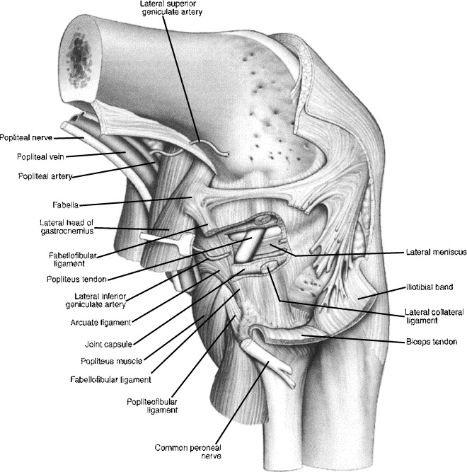
I've got 2 patients in orbit with concomitant fibula fractures (PLC injuries) in context of tibial plateau fracture. PLC anatomy does my head in, so heres a list of what im reading. If anyone has some tips on management of these, please chime in! #orthotwitter
and whilst i agree neck injuries heal (and rarely need to be actively managed) im concerned about these 2 specific cases in that the PLC is identifiably disrupted as a descrete structure/complex, and a posterolateral instabilty ist quite likely (though not testable at present)
Radial N revision follow exfix of a dist humerus fracture. Radial paresis/paraesthesia. Nerve found entirely intact, and wasn’t wrapped around the exfix pin visible immediately deep to it. The nerve turned a hard corner over the pin in flexion, hopefully it wakes up #orthotwitter
Dorsal lateral paratricipital window. Opened the lateral intermuscular septum. Humeral cortex to the right. Blue are the exfix pins. Yellow is the radial nerve. Green is the posterior cutaneous antebrachial nerve. @kangsta77@Gnomelover1970
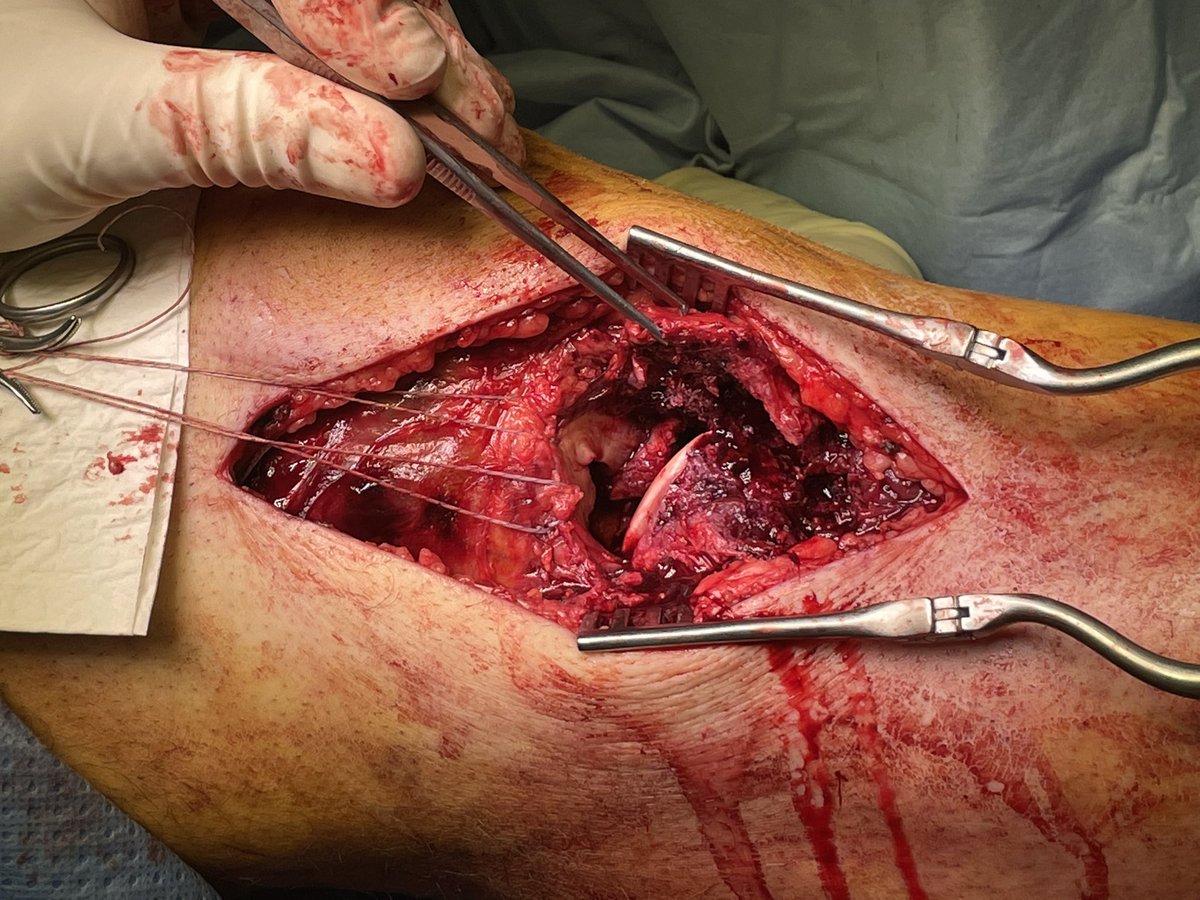
Quick n dirty no frills lateral tibia walkthrough. Longitudinal lateral/anterolateral extensile approach for more complex fracture constellations requiring open reduction. The big brother of the Lazy S /MIPO approach #orthotwitter
Landmarks and positioning
Probably didn’t really need to extend proximally to the lateral epicobdyle, could’ve saved a cm or 2
Part 1 of a trilogy! Reconstructing a Moore 2 Schatzker IV fracture dislocation. Middle aged fall from a ladder. I’ll start with first and finals, then start a new thread with progressive intraop fluros, and lastly a third thread with clinical anatomy photos #orthotwitter
Pre and post op CT forensics #1:
Coronal slice posterior third of the plateau. Not going to pretend the lateral joint surface is perfect, looks oddly wide, though condylar with is spot on. direct arthrotomy certainly looked pretty good. Don’t know where that crumb came from. Grrr
Pre and post op CT forensics #2:
Coronal slice anterior third of the joint. That impacted lat fragment belonged post and is seen reduced in the previous image. Here the ant lat joint block was ok-ish just that defect in transition into eminentia. Medial was a mess (comes later!)