William Aird Profile picture
Feb 1, 2023 4 tweets 2 min read Read on X
1/4

#GI #GIbleed #urea

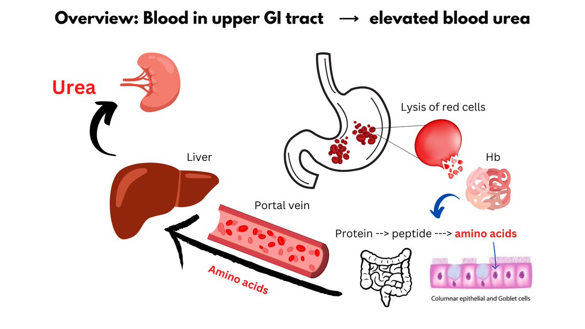
Elevated BUN: creatinine ratio favors UPPER over lower GI bleed.

Why?

1) Hb in stomach --> amino acids (aa).
2) aa absorbed by small intestine.
3) aa transported and taken up by liver.
4) In the liver, aa undergo deamination, leading to release of NH3. Image
2/4

5) NH3 (aka ammonia) is converted to urea by the urea cycle, leading to elevated blood urea levels.
6) Meantime, creatinine is not influenced by the protein/aa bolus.
7) Thus, BUN/creatinine ratio is increased.
3/4

So why does blood cause this bump in urea and increase in BUN/creatinine ratio, whereas a steak dinner does not?

Because blood has poor BIOLOGICAL VALIUE (the protein is poorly utilized by the body).

The LOWER the biological value of a protein, the HIGHER the urea.
4/4

For more information see:

thebloodproject.com/cases-archive/…

• • •

Missing some Tweet in this thread? You can try to force a refresh
 

Keep Current with William Aird

William Aird Profile picture

Stay in touch and get notified when new unrolls are available from this author!

Read all threads

This Thread may be Removed Anytime!

PDF

Twitter may remove this content at anytime! Save it as PDF for later use!

Try unrolling a thread yourself!

how to unroll video
  1. Follow @ThreadReaderApp to mention us!

  2. From a Twitter thread mention us with a keyword "unroll"
@threadreaderapp unroll

Practice here first or read more on our help page!

More from @WilliamAird4

Sep 15
1/7

PSEUDOTHROMBOCYTOPENIA

I tweeted a poll asking for the next step in a patient with thrombocytopenia and a platelet clumping on a peripheral smear. 62% of you answered correctly, namely to repeat the CBC in a green top (heparin-containing) tube. Image
2/7

Key points:

1) Pseudothrombocytopenia is mediated by EDTA-dependent antibodies that react with platelets in blood that is anticoagulated with EDTA (chelates calcium, necessary for clotting reactions), causing platelet clumping and falsely low platelet counts.
3/7

2) Pseudothrombocytopenia is purely an in vitro artifact.

3) EDTA-containing collection tubes are preferred for CBCs/peripheral smears because, compared with other anticoagulants, EDTA allows the best preservation of cellular components and blood cell morphology
Read 7 tweets
Jul 22
1/6

LYMPHOCYTOPENIA (aka lymphopenia)

A) Definition

Absolute L count < 1,000/microliter or 1.0 x 10^9/L Image
2/6

B) Causes Image
3/6

C) Mechanisms

i. Reduced production
ii. Increased destruction
iii. Increased apoptosis
iv. Redistribution blood to lymphoid tissue Image
Read 6 tweets
Jun 11
1/7

RELATIONSHIPS BETWEEN RBC INDICES

RBC count

a. Used to calculate the Hct (Hct = MCV x RBC count)
b. Used by some to predict iron deficiency vs. thalassemia (e.g., Meltzer index)
b. Largely ignored because it says nothing about the size/Hbization of RBCs Image
2/7

... you could have LOTS of SMALL RBCs or FEWER LARGE RBCs amounting to the same Hct (viscosity) and Hb (oxygen carrying capacity)!
3/7

MCV

a. Ah, now we're talking 😀! MCV is super helpful because it is the first branch point in the ddx of anemia - microcytic vs. normocytic vs. macrocytic. Each has its own specific causes.
Read 7 tweets
May 31
1/4

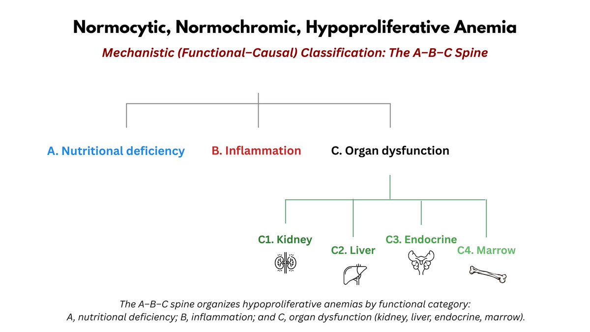
APPROACH TO NORMOCYTIC ANEMIA

This is the most common type of anemia, and its differential diagnosis can sometimes feel overwhelming, and 'all over the place'.

The following is organizational scheme with simple diagnostic buckets that covers virtually all causes: Image
2/4

BUCKET LIST 1:

Appropriate vs. inappropriate retic response (appropriate defined by absolute retic count > 120 x 10^9/L or 0.12 x 10^12/L)?

BUCKET LIST 2:

If appropriate retic response, is there bleeding or hemolysis? Image
3/4

BUCKET LIST 3:

If inappropriate retic response, is there inflammation or organ dysfunction?

BUCKET LIST 4:

If organ dysfunction, is there chronic liver disease, chronic kidney disease, endocrinopathy or bone marrow dysfunction?
Read 4 tweets
May 30
1/5

COAGULATION IN VITRO/IN VIVO

In vitro (test tube):

1. The clotting cascade in plasma is activated by the addition of a negative charge, which activates the intrinsic pathway (aPTT) or tissue factor, which activates the extrinsic pathway (PT) in presence of Ca2+ and PL. Image
2/5

2. Once the ingredients are added together, the "stop watch" is started and time to clot formation at 37C is measured (the assays do not discriminate between crosslinked and uncrosslinked fibrin).
3/5

In vivo (the body):

1. The clotting cascade is ALWAYS activated by TF-mediated activation of FVII. TF hides in the subendothelial layer of the blood vessel wall and is expressed on the surface of activated monocytes.
Read 5 tweets
May 21
1/6

NORMOCYTIC, NORMOCHROMIC IRON DEFICIENCY ANEMIA (IDA)

We tend to think of IDA as being microcytic (more consistently than hypochromic). However, there are certain situations in which the MCV is normal in IDA. Image
2/6

Examples include:

1. A patient with high-normal baseline MCV whose MCV falls within the normal range in IDA (see first graphic for an example).

2. A patient with baseline macrocytosis, for example from concomitant liver disease, B12 deficiency, MDS or hydroxyurea use.
3/6

3. Rare cases where the MCV simply doesn't budge from baseline despite clear cut evidence of IDA (i.e. anemia +/- low MCHC with low ferritin that responds to iron treatment).
Read 6 tweets

Did Thread Reader help you today?

Support us! We are indie developers!


This site is made by just two indie developers on a laptop doing marketing, support and development! Read more about the story.

Become a Premium Member ($3/month or $30/year) and get exclusive features!

Become Premium

Don't want to be a Premium member but still want to support us?

Make a small donation by buying us coffee ($5) or help with server cost ($10)

Donate via Paypal

Or Donate anonymously using crypto!

Ethereum

0xfe58350B80634f60Fa6Dc149a72b4DFbc17D341E copy

Bitcoin

3ATGMxNzCUFzxpMCHL5sWSt4DVtS8UqXpi copy

Thank you for your support!

Follow Us!

:(