🧵🪡A thread on our paper out in today’s @Dev_Cell issue, featuring one of my images on the cover 🤩 

You might’ve heard about the #BBB but have you heard about the BNB (Blood Nerve Barrier)? #pns #bloodnervebarrier 

We showed that endoneurial blood vessels (EndoBVs) are surrounded by pericytes (alphaSMA, orange) 🤗 @ImarisSoftware
And another component of the vascular unit being tactocytes (green, with characteristic rER) in close contact with the basement membrane of EndoBVs 

The last cell of the vascular unit of the BNB are macrophages (Iba1 or F4/80, blue) 🐭 @ImarisSoftware
We found that the VU is conserved in rat 🐀 and human 👩🏽🤝👨🏼samples. Human samples shown here: beautiful #vEM z slice from @EM__Ian with characteristic endothelial cell/#pericyte peg-socket interactions & @ImarisSoftware of tactocytes and macrophages along EndoBVs 

Next, we looked at how the BNB is conferred and showed that EndoBVs have specialised tight junctions and low levels of transcytosis. 


We then used our P0-RafTR model (previously published @NeuroCellPress) where we could “open” the BNB in a regulated fasion. Here, i.v Evans Blue 🔵 

And we found that opening the BNB is by strongly increased transcytosis (plvap), while tight junctions (here #claudin5) were maintained. 

Interestingly, Liza found that #Macrophages were the only cells taking up tracers (here HRP) and that they move away from the BNB in the P0-RafTR model. In short, acting as a secondary barrier mechanism. 

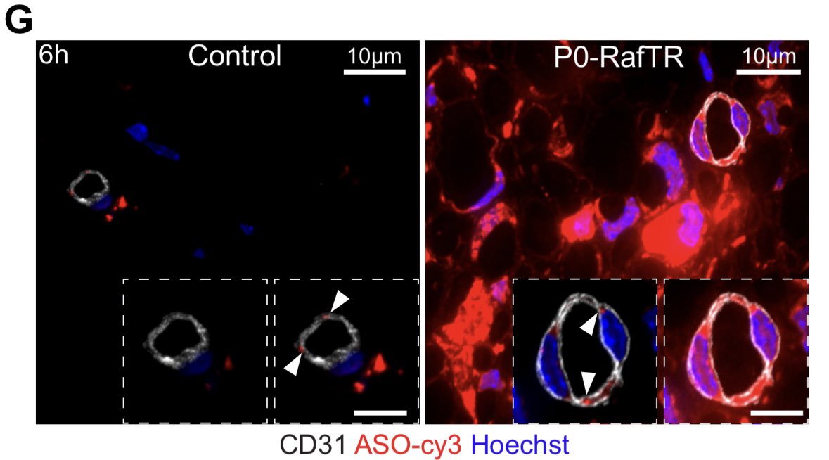
Finally, we looked at the therapeutic potential in collaboration with @ionispharma and showed increased antisense oligos (ASOs) inside endoneurial endothelial cells & the endoneurium when we open the BNB as well as significantly increased target gene downregulation. 


So much more in the paper! Check it out and tell us what you think!
Most importantly, thank you to everyone involved! Especially Liza, @EM__Ian @JemimaBurden @AnnelaureCattin & many more not on Twitter! The animals 🐭& staff looking after them, the @LMCB_UCL staff which keeps everything running 🌟 and our funders @CRUKresearch @wellcometrust
This is the link: sciencedirect.com/science/articl…
• • •
Missing some Tweet in this thread? You can try to
force a refresh


