Sign?
Diagnosis?
Diagnosis?

Answer
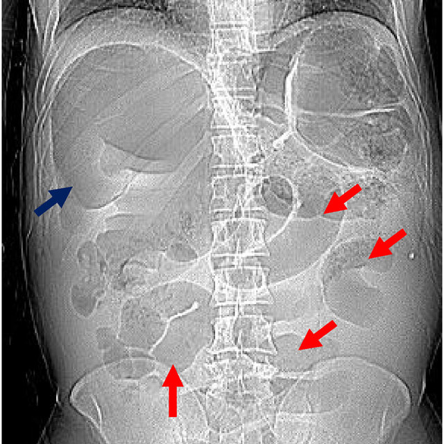
Football Sign
Football Sign
Lucencies show outline of the falciform ligament showing large pneumoperitoneum (in this case a pediatric patient).
If you see the football sign, think free air!
If you see the football sign, think free air!

Football sign (pneumoperitoneum)
👉massive pneumoperitoneum, where the abdominal cavity is outlined by gas from a perforated viscus
👉 median umbilical ligament and falciform ligament are sometimes included in the description of this sign, as representing the sutures.
👉massive pneumoperitoneum, where the abdominal cavity is outlined by gas from a perforated viscus
👉 median umbilical ligament and falciform ligament are sometimes included in the description of this sign, as representing the sutures.

Which football is used as the model ball varies according to the nationality of the author.
Rugby, Australian rules, and American football all have balls that fit the bill. Soccer is clearly not appropriate.
Rugby, Australian rules, and American football all have balls that fit the bill. Soccer is clearly not appropriate.
It is most frequently seen in children with advanced necrotizing enterocolitis.
Other causes
bowel obstruction with secondary perforation
malrotation with midgut volvulus
Hirschsprung disease
meconium ileus
intestinal atresia
peptic ulcer disease
iatrogenic
endoscopic perf
Other causes
bowel obstruction with secondary perforation
malrotation with midgut volvulus
Hirschsprung disease
meconium ileus
intestinal atresia
peptic ulcer disease
iatrogenic
endoscopic perf
• • •
Missing some Tweet in this thread? You can try to
force a refresh