#POCUS question of the day?
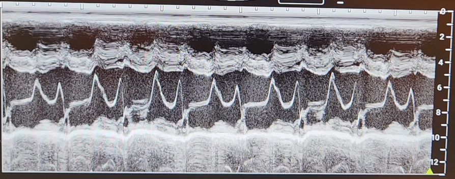
1. What does this image show, and what mode of ultrasound was used to obtain it?
2. What is the E-point?
3. What is the A- point?
4. What is EPSS, and why do we use it?
Scroll down to learn how to interpret the image.
1. What does this image show, and what mode of ultrasound was used to obtain it?
2. What is the E-point?
3. What is the A- point?
4. What is EPSS, and why do we use it?
Scroll down to learn how to interpret the image.

Answer 1: The image above shows the mitral valve in M mode taken during the PLAX view at the MV leaflet tips.
Answer 2: During early diastole, the leaflets separate widely with the maximum early diastolic motion of the anterior leaflet termed the E-point
Answer 2: During early diastole, the leaflets separate widely with the maximum early diastolic motion of the anterior leaflet termed the E-point

Answer 3: During atrial contraction, the leaflets move towards each other in mid-diastole (F-point =diastasis) and then separate again with atrial systole, thus resulting in the late diastolic peak or the A point.
Extra Credit
F Point: marks the onset of diastasis slow flow
C Point: The point where the anterior and posterior leaflets of the mitral valve come together during systole.
D Point: Mitral valve opening at the beginning of diastole
F Point: marks the onset of diastasis slow flow
C Point: The point where the anterior and posterior leaflets of the mitral valve come together during systole.
D Point: Mitral valve opening at the beginning of diastole

Answer 4: EPSS = E-point septal separation and is defined as the distance between the anterior leaflet of the mitral valve and the interventricular septum. 

• • •
Missing some Tweet in this thread? You can try to
force a refresh


