QUESTION: Do either/both of these patients have anisocytosis?
NOTE: RDW-CV and RDW-SD are discordant.
2/5
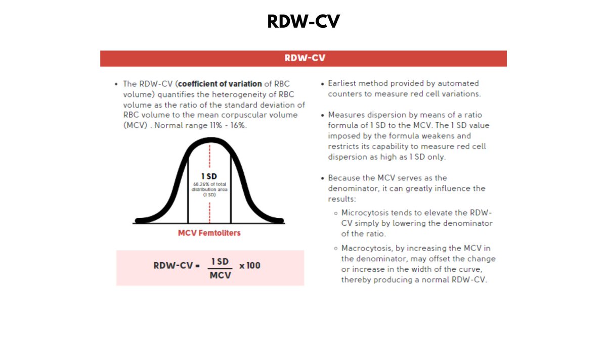
MCV represents the average volume of RBCs measured, but does not provide information about variation in cell size.
The latter can be estimated by measuring the diameter of many RBCs under the light microscope. More commonly it is generated as the RDW by automated counters.
3/5
2 ways to express the RDW:
1. RDW-CV is a percentage of the SD/MCV, thus the lower the MCV, the higher the RDW-CV. Conversely, the higher the MCV, the lower the RDW-CV.
4/5
2. The RDW-SD is a direct measurement of variation in cell volume, and is not influenced by cell size. Thus, it is a better marker for anisocytosis.
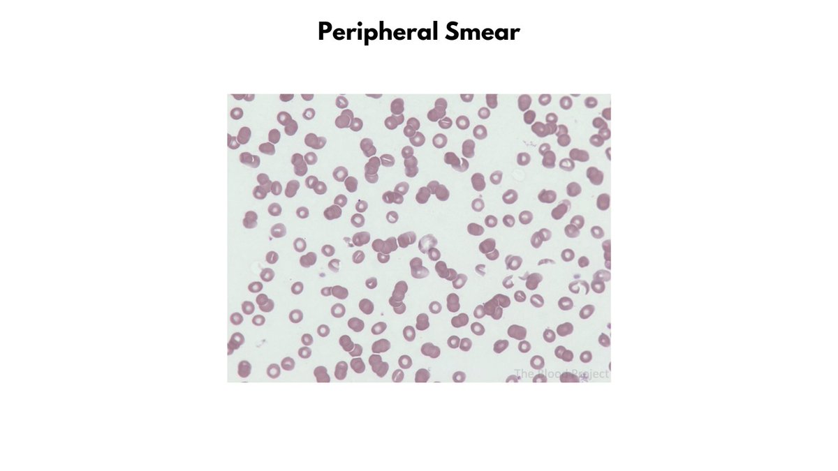
Patient A does not have anisocytosis despite the high RDW-CV.
Patient B has anisocytosis despite the normal RDW-CV.
5/5
EASY TO REMEMBER:
a. RDW-SD > 46 fL = anisocytosis
b. A lower than normal RDW-SD is clinically meaningless - lower values reflect less biological noise.
67 yo M with history of colon cancer presents to ED feeling unwell. He is disoriented with T 102F, BP 70/40, HR 120/min. Labs are shown in graphic. He is intubated, treated with fluids/pressors and Abx, and passes away 18 h later.
What stands out in the labs?
2/8
Labs show:
1. Profound reduction in Hb over 11 h 2. All blood samples hemolyzed 3. Pseudohyperkalemia (from hemolysis) 4. Positive hemolytic makers EXCEPT NORMAL HAPTOGLOBIN
... Hmm, but haptoglobin is the most sensitive marker of hemolysis!
3/8
This was a case of clostridium perfringens sepsis, which is almost universally fatal. It occurs primarily in immunocompromised patients and usually arises from the GI tract.
C. perfringens is a cause of (massive) NON-IMMUNE, EXTRA-CORPUSCULAR HEMOLYSIS.
IRON DEFICIENCY ANEMIA (IDA) vs. THALASSEMIA (THAL) TRAIT
Take a look at the two CBCs in the graphic and identify which belongs to a patient with IDA (ferritin < 10), and which belongs to the individual with beta-thal trait (increased HbA2).
2/6
The CBC on top is from a person with beta thal, the one on the bottom from a patient with IDA.
Since 1973, several formulas have been established as simple, fast, and inexpensive tools to differentiate β-thalassemia (β-thal) trait from iron deficiency anemia (graphic).
3/6
None of the formulas is highly specific or sensitive (which is why there are so many options!).
Let's see how the various formulas fare in this case, beginning with the Mentzer index - perhaps the most popular of the discriminant formulas.
Most mammals share the same clotting cascade. But there are exceptions. Check out the clotting assays from dolphins in the graphic. Note that their PT is comparable to humans, but they do not clot in the aPTT assay.
Q: What are possible explanations for this finding?
2/6
ANSWER:
Deficiency of or (much less likely) inhibitor against one or more components of the intrinsic pathway, which is assayed by the aPTT and includes FXII, FXI, FIX, and FVIII.
3/6
A 1969 paper in Science showed that dolphins and killer whales had complete absence of functional FXII, explaining their prolonged aPTT.
Authors: "The body has some other means by which it can activate coagulation than through the intervention of activated factor XII".
The Ancient Greeks codified bloodletting as a rational therapy for disease, based on the theory of the humors. Health reflected the balance and free flow of the humors and their qualities, while disease arose from imbalances.
2/8
Treatment was aimed toward helping Nature restore the humors to their proper equipoise. One such strategy was to remove blood when it was in excess or corrupt.
Galen (129-216 AD) was an ardent bloodletter. He would often draw off blood until his patient fainted.
3/8
In his Method of Healing, he wrote:
"I carefully drew enough [blood] from him that he fainted, having learned from reason and experience that this is the best remedy for continual fevers when [the patient’s] strength is vigorous..."
74 yo F presents with CBC shown in graphic below. You will note she has macrocytic anemia, which has a wide differential diagnosis (also shown in graphic).
2/11
The patient's reticulocyte count was elevated (not shown), possibly accounting for the macrocytosis and pointing to presence of either bleeding or hemolysis.
Hemolysis labs were in fact positive (see graphic below). What is missing in the hemolysis labs?
3/11
The AST:ALT ratio, which was elevated in this case, and demonstration of positive blood in urine by dipstick in absence of red blood cells under the microscope (i.e., evidence of hemoglobinuria).