Let me take you on a journey... #orthotwitter
"no, the triceps tendon was not augmented, why do you ask?"
no problem, we'll revise it, this time with augmentation.
yeah, no.
I got to revise it after the 2nd failure. went with 2 stage just to make sure (took the short 72hr microbiology option). took everything out, Joint absolutley stable through full PROM. ABx spacer.
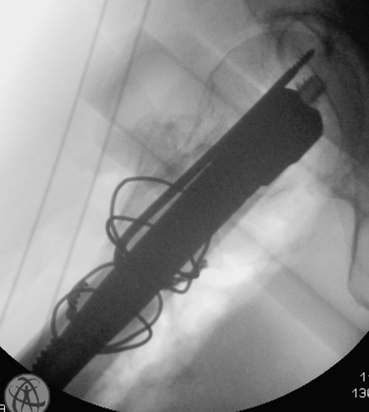
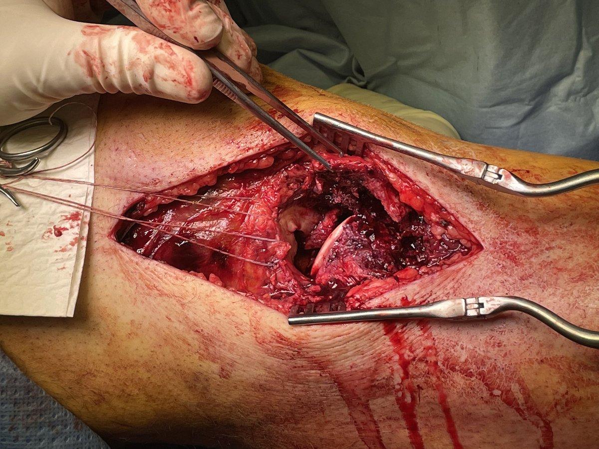
Given joint stability, aim was to readapt tricpes for extensor function. revised at 5 days, no indication of infection. 4 Strand fibertape locking stitch into triceps (mobilised and distalised a bit) then ran fibertape through the fracture and intramedullary down the ulnashaft,… twitter.com/i/web/status/1…
moral of the story - suture augment the triceps tendon during the index surgery!

• • •

Missing some Tweet in this thread? You can try to force a refresh
 

Keep Current with Dr. Edward Oates

Dr. Edward Oates Profile picture

Stay in touch and get notified when new unrolls are available from this author!

Read all threads

This Thread may be Removed Anytime!

PDF

Twitter may remove this content at anytime! Save it as PDF for later use!

Try unrolling a thread yourself!

how to unroll video
  1. Follow @ThreadReaderApp to mention us!

  2. From a Twitter thread mention us with a keyword "unroll"
@threadreaderapp unroll

Practice here first or read more on our help page!

More from @traumaticum

Mar 20
Some days i regret getting out of bed
#orthotwitter ImageImage
I'll post these, even though theyre far from pretty. lots of reasons why this or that shouldve couldve wouldve. not something i enjoyed much. Cables doing their best where there was no tube to reconstruct. 85yo bone was dust. screw went into the best bit of the head. open to… twitter.com/i/web/status/1… ImageImage
both vastus lat and glut med on the trochanter fragment. made getting past the lateral wall of tissue a huge PITA without defunctioning something big. managed tho. dozens of intermediate fragnment. only read was lateral distal trochanter to diaphysis. only had 10mm of overlap.
Read 5 tweets
Nov 20, 2022
Cerclage wires got a good wrap in 2022. Several conference presentations, a couple of posters and abstracts, and now here's a mini literature collection for @DrMarecek and myself to bath in the next time the topic comes up #orthotwitter

osteosynthesis.org/42003807-commi…
just an aside, i was honestly a bit skeptical whether people would follow links to off-site content, and looking at the 30 odd likes on this tweet i was thinking "well there you go, not much interest/engagement". huh.. was i wrong, i just looked at site stats ~600 click-throughs!
Read 4 tweets
May 6, 2022
I've got 2 patients in orbit with concomitant fibula fractures (PLC injuries) in context of tibial plateau fracture. PLC anatomy does my head in, so heres a list of what im reading. If anyone has some tips on management of these, please chime in! #orthotwitter
and whilst i agree neck injuries heal (and rarely need to be actively managed) im concerned about these 2 specific cases in that the PLC is identifiably disrupted as a descrete structure/complex, and a posterolateral instabilty ist quite likely (though not testable at present)
Read 8 tweets
Mar 16, 2022
Radial N revision follow exfix of a dist humerus fracture. Radial paresis/paraesthesia. Nerve found entirely intact, and wasn’t wrapped around the exfix pin visible immediately deep to it. The nerve turned a hard corner over the pin in flexion, hopefully it wakes up #orthotwitter Image
Dorsal lateral paratricipital window. Opened the lateral intermuscular septum. Humeral cortex to the right. Blue are the exfix pins. Yellow is the radial nerve. Green is the posterior cutaneous antebrachial nerve. @kangsta77 @Gnomelover1970 Image
Just testing this out

@threadreaderapp unroll
Read 4 tweets
Feb 14, 2022
Quick n dirty no frills lateral tibia walkthrough. Longitudinal lateral/anterolateral extensile approach for more complex fracture constellations requiring open reduction. The big brother of the Lazy S /MIPO approach #orthotwitter Image
Landmarks and positioning ImageImage
Probably didn’t really need to extend proximally to the lateral epicobdyle, could’ve saved a cm or 2 ImageImage
Read 12 tweets
Feb 12, 2022
Part 1 of a trilogy! Reconstructing a Moore 2 Schatzker IV fracture dislocation. Middle aged fall from a ladder. I’ll start with first and finals, then start a new thread with progressive intraop fluros, and lastly a third thread with clinical anatomy photos #orthotwitter ImageImage
Pre and post op CT forensics #1:
Coronal slice posterior third of the plateau. Not going to pretend the lateral joint surface is perfect, looks oddly wide, though condylar with is spot on. direct arthrotomy certainly looked pretty good. Don’t know where that crumb came from. Grrr ImageImage
Pre and post op CT forensics #2:
Coronal slice anterior third of the joint. That impacted lat fragment belonged post and is seen reduced in the previous image. Here the ant lat joint block was ok-ish just that defect in transition into eminentia. Medial was a mess (comes later!) ImageImage
Read 8 tweets

Did Thread Reader help you today?

Support us! We are indie developers!


This site is made by just two indie developers on a laptop doing marketing, support and development! Read more about the story.

Become a Premium Member ($3/month or $30/year) and get exclusive features!

Become Premium

Don't want to be a Premium member but still want to support us?

Make a small donation by buying us coffee ($5) or help with server cost ($10)

Donate via Paypal

Or Donate anonymously using crypto!

Ethereum

0xfe58350B80634f60Fa6Dc149a72b4DFbc17D341E copy

Bitcoin

3ATGMxNzCUFzxpMCHL5sWSt4DVtS8UqXpi copy

Thank you for your support!

Follow Us on Twitter!

:(