William Aird Profile picture
Jul 1, 2023 9 tweets 3 min read Read on X
1/9

EXAMPLE OF Hb-Hct DISCORDANCE

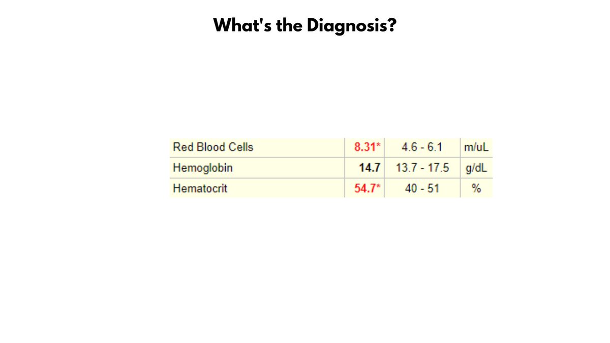
Take a look at the values in the graphic. Without looking ahead in the thread, can you make a diagnosis?
2/9

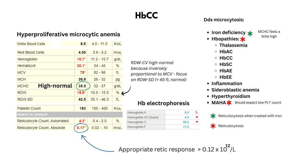
The patient has ERYTHROCYTOSIS (defined as an increased RBC count) and POLYCYTHEMIA (based on the Hct, but not the Hb).
3/9

What's up with the elevated Hct but normal Hb? Well, there is violation of the 3:1 rule which states that the Hct is normally 3x the Hb (e.g., Hct 45, Hb 15). Stated another way, the MCHC (Hb/Hct) is low, thus there is HYPOCHROMIA.
4/9

We can calculate not only the MCHC but also the MCV (Hct/RBC count). The MCV is only 66 fL, so the patient has non-anemic hypochromic microcytosis.
5/9

To summarize, the patient has erythrocytosis, polycythemia (according to Hct), and non-anemic hypochromic microcytosis.
6/9

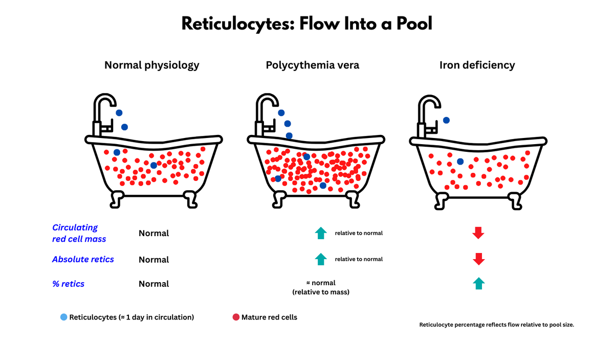
We can illustrate the RBCs using a schematic of spun hematocrits.
7/9

This is a patient with newly diagnosed Jak2 V617F-positive polycythemia vera. Many such patients present with concomitant iron deficiency (from increased Fe demand +/- occult GI bleeding).
8/9

It's really interesting to consider that the patient with PV may present with erythrocytosis alone (masked PV), then develop erythrocytosis and polycythemia (+/- iron deficiency) and finally - during a specific window of treatment - polycythemia alone. So cool!!
9/9

When considering the Hb and Hct in polycythemia vera, the Hct is the critical parameter because it is the primary determinant of blood viscosity and thrombotic complications. So, the presence of a normal Hb - as in this case - should not provide reassurance!

• • •

Missing some Tweet in this thread? You can try to force a refresh
 

Keep Current with William Aird

William Aird Profile picture

Stay in touch and get notified when new unrolls are available from this author!

Read all threads

This Thread may be Removed Anytime!

PDF

Twitter may remove this content at anytime! Save it as PDF for later use!

Try unrolling a thread yourself!

how to unroll video
  1. Follow @ThreadReaderApp to mention us!

  2. From a Twitter thread mention us with a keyword "unroll"
@threadreaderapp unroll

Practice here first or read more on our help page!

More from @WilliamAird4

Oct 21
1/7

A STRUCTURED APPROACH TO NORMOCYTIC ANEMIA

A practical, population, and molecular look at how we reason through normocytic hypoproliferative anemia at the bedside.

(Full TBP tutorial linked below ⬇️)
2/7

1️⃣ The ABC Spine – A Bedside Framework

A: substrate supply (nutritional)
B: regulatory signaling (inflammatory)
C: organ dysfunction (kidney, liver, endocrine, marrow)

We focus here on the hypoproliferative end of the spectrum, where the marrow is not keeping up. Image
3/7

2️⃣ Anemia of the Elderly – A Population Perspective

In older adults, causes divide roughly:

1/3 nutritional 
1/3 inflammatory
1/3 unexplained

Each slice maps back to A, B, or C of the spine. Image
Read 7 tweets
Oct 7
IV IRON AND FERRITIN

1/8

I recently posted a question about the spike in serum ferritin after IV iron.

It can shoot from <10 to >1000, yet transferrin saturation (TSAT) only rises to normal — it barely moves.

What’s going on?

This is macrophage physiology in action 👇 Image
2/8

Step 1 – Uptake

IV iron–carbohydrate complexes (e.g., iron dextran) are endocytosed by macrophages in the liver, spleen, and marrow.

Inside, the iron is released and floods the labile Fe²⁺ pool, the cell’s internal “iron bank.”
3/8

Step 2 – Ferritin synthesis explodes

High intracellular Fe²⁺ flips the IRP–IRE switch, unleashing translational ferritin synthesis.

Cytokines add a transcriptional boost.

Ferritin production ↑↑ → serum ferritin rises via a regulated, non-classical secretion pathway.
Read 8 tweets
Oct 6
1/4

Patients with SLE can present with a staggering array of hematologic abnormalities. Virtually no component of the blood is spared. Everything is fair game:

1. Blood cells - cytopenia and cytoses
2. Hemostasis - thrombosis, TMA
3. Lymphadenopathy/splenomegaly
4. Lymphoma Image
2/4

Diagnostic criteria for SLE include the following hematological parameters:

1. Hemolytic anemia
2, Leukopenia
3. Lymphopenia
4. Thrombocytopenia
5. Anti phospholipid antibodies
3/4

The primary mechanisms of hematological alterations in SLE are:

1. Autoimmunity
2. Immunosuppressive medications

There is increasing appreciation that the disease targets not only peripheral blood cells but also the bone marrow leading to:

1. MF
2. PRCA
3. AA Image
Read 4 tweets
Sep 20
1/4

The Plasma Exchange Paradox

Have you ever wondered why plasma exchange saves lives in TTP but does nothing in ITP? Both are IgG-mediated. So why the difference?
2/4

In ITP, antibodies opsonize platelets. Even if you remove some by exchange, IgG quickly re-equilibrates from the extravascular pool and continues to be produced. With no missing factor to replace, there’s no lasting benefit.
3/4

In TTP, antibodies inhibit ADAMTS13. Plasma exchange not only removes antibody but also replaces the missing enzyme (and helps clear ULVWF multimers). That dual action is why it works.
Read 4 tweets
Aug 26
1/5

SPLENOMEGALY/HYPERSPLENISM

Mechanisms of splenomegaly:
• Congestive
• Work hypertrophy
• Infiltrative

What enlarges?
• Red pulp: macrophages, sinusoids, blood
• White pulp: lymphocytes, plasma cells
• Infiltrative: tumor, storage cells, granulomas, amyloid

#MedEd Image
2/5

SPLENOMEGALY/HYPERSPLENISM

Red pulp = ~75% of spleen, the “filter & reservoir.”

Site of pooling, culling & destruction → main driver of splenomegaly + hypersplenism.

Open circulation = quality control: normal cells pass, rigid ones get removed. Image
3/5

Splenomegaly → hypersplenism when an enlarged spleen starts causing cytopenias:

• Pooling (cells trapped in red pulp)
• Destruction (macrophage culling)

→ ↓ platelets>WBCs>RBCs

All hypersplenism comes with splenomegaly, but not all splenomegaly causes hypersplenism Image
Read 5 tweets
Jan 19
1/7

I posted the graphic below earlier in the week and asked what was missing.

Before addressing the question, let's flesh out the various diagnostic containers.

1. HEMOLYSIS:

Immune hemolysis:
Autoimmune
Warm, Cold, Mixed
Alloimmune
ATR, DTR Image
2/7

Non-immune hemolysis
Intracapsular
Hemoglobinopathies
Membrane disorders
Hbopathies

Extracorpuscular
Infection (babesiosis, malaria, clostridial)
Liver (spur cell anemia, Zieve syndrome)
TMA
3/7

Non-immune hemolysis (cont'd)
Extracorpuscular
Valve hemolysis
March hemoglobinuria
Venom
Hyperthermia

2. BLEEDING:

External:
Hemorrhage
Phlebotomy
Internal (hematoma)
Read 7 tweets

Did Thread Reader help you today?

Support us! We are indie developers!


This site is made by just two indie developers on a laptop doing marketing, support and development! Read more about the story.

Become a Premium Member ($3/month or $30/year) and get exclusive features!

Become Premium

Don't want to be a Premium member but still want to support us?

Make a small donation by buying us coffee ($5) or help with server cost ($10)

Donate via Paypal

Or Donate anonymously using crypto!

Ethereum

0xfe58350B80634f60Fa6Dc149a72b4DFbc17D341E copy

Bitcoin

3ATGMxNzCUFzxpMCHL5sWSt4DVtS8UqXpi copy

Thank you for your support!

Follow Us!

:(