William Aird Profile picture
Jul 1 9 tweets 3 min read Twitter logo Read on Twitter
1/9

EXAMPLE OF Hb-Hct DISCORDANCE

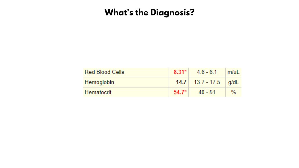
Take a look at the values in the graphic. Without looking ahead in the thread, can you make a diagnosis?
2/9

The patient has ERYTHROCYTOSIS (defined as an increased RBC count) and POLYCYTHEMIA (based on the Hct, but not the Hb).
3/9

What's up with the elevated Hct but normal Hb? Well, there is violation of the 3:1 rule which states that the Hct is normally 3x the Hb (e.g., Hct 45, Hb 15). Stated another way, the MCHC (Hb/Hct) is low, thus there is HYPOCHROMIA.
4/9

We can calculate not only the MCHC but also the MCV (Hct/RBC count). The MCV is only 66 fL, so the patient has non-anemic hypochromic microcytosis.
5/9

To summarize, the patient has erythrocytosis, polycythemia (according to Hct), and non-anemic hypochromic microcytosis.
6/9

We can illustrate the RBCs using a schematic of spun hematocrits.
7/9

This is a patient with newly diagnosed Jak2 V617F-positive polycythemia vera. Many such patients present with concomitant iron deficiency (from increased Fe demand +/- occult GI bleeding).
8/9

It's really interesting to consider that the patient with PV may present with erythrocytosis alone (masked PV), then develop erythrocytosis and polycythemia (+/- iron deficiency) and finally - during a specific window of treatment - polycythemia alone. So cool!!
9/9

When considering the Hb and Hct in polycythemia vera, the Hct is the critical parameter because it is the primary determinant of blood viscosity and thrombotic complications. So, the presence of a normal Hb - as in this case - should not provide reassurance!

• • •

Missing some Tweet in this thread? You can try to force a refresh
 

Keep Current with William Aird

William Aird Profile picture

Stay in touch and get notified when new unrolls are available from this author!

Read all threads

This Thread may be Removed Anytime!

PDF

Twitter may remove this content at anytime! Save it as PDF for later use!

Try unrolling a thread yourself!

how to unroll video
  1. Follow @ThreadReaderApp to mention us!

  2. From a Twitter thread mention us with a keyword "unroll"
@threadreaderapp unroll

Practice here first or read more on our help page!

More from @WilliamAird4

Jul 2
1/4

DOHLE BODY vs. MAY-HEGGLIN INCLUSION

Döhle bodies and May-Hegglin inclusions (also called Döhle-like bodies) are distinct entities. They both stain sky blue on peripheral smears, and they are both found in neutrophils, but the similarities stop there.
2/4

Döhle bodies are acquired, transient, associated with sepsis and burns, and appear as poorly defined blue inclusions often at the periphery of neutrophils, bands and metamyelocytes. EM studies show that they contain stacks of rough endoplasmic reticulum.
3/4

May-Hegglin inclusions are found in patients with MYH9-related disease (RD) which is caused by mutations in the myosin-9 gene and associated with thrombocytopenia and giant platelets, hearing loss, cataracts and nephropathy.
Read 4 tweets
Jun 30
1/6

HLH DIAGNOSIS

Hemophagocytic lymphohistiocytosis (HLH) is a rare, life-threatening hyperinflammatory syndrome of intense immune activation associated with fever, hepatosplenomegaly, cytopenias, hyperferritinemia, and presence of activated macrophages in hemopoietic organs.
2/6

HLH in adults is usually initiated by external causative factors such as drugs and infections or in the context of certain underlying diseases including cancer or autoimmune disease.
3/6

Diagnosis of HLH relies on clinical scoring systems since there no single laboratory finding has the sensitivity and specificity to definitively detect HLH. There are two commonly employed scoring systems:

1. HLH-2004 criteria
2. HScore
Read 6 tweets
Jun 29
1/5

HbSC

I have seen a number of patients with HbSC disease lately, and thought it would be helpful to review the differences between HbSC and HbSS.

1. HbSC disease is caused by co-inheritance of the hemoglobin S (HbS) and hemoglobin C (HbC) beta globin gene mutations.
2/5

2. HbC is the second most common hemoglobin variant after HbS in the US, and third most common worldwide after HbS and HbE. HbC Confers protection against severe malaria.

3. HbSC accounts for 30% of sickle cell disease (SCD) in the US and UK.
3/5

4. Pathophysiology of HbSC disease involves:

1) RBC dehydration leading to increased HbS concentration and sickling under deoxygenated conditions.

2) HbC crystallization under oxygenated conditions.

3) Increased blood viscosity.
Read 5 tweets
Jun 28
1/9

VITAMIN K DEFICIENCY

I recently saw a 34 yo F with a history of radiologically and biopsy-proven primary sclerosing cholangitis who was found to have an elevated PT/INR and aPTT (no bleeding symptoms, no warfarin/antibiotics). Her vitamin A, D and E levels were all low.
2/9

She was treated with vitamin K 5 mg IV x 2, and her INR normalized within 12 hours. Based on her low levels of vitamin A, D and E she was diagnosed with primary sclerosing cholangitis-associated fat-soluble vitamin deficiency.
3/9

Q: WHAT IS PRIMARY SCLEROSING CHOLANGITIS (PBC)?

A: Chronic progressive liver disease where inflammation and fibrosis lead to multifocal biliary strictures.
Read 9 tweets
Jun 1
1/4

I tweeted a CBC time series yesterday with questions. The answers are shown in the graphic.

This was a young F with reactive thrombocytosis secondary to tubo-ovarian abscess. She was treated with surgical drainage and antibiotics with resolution of her thrombocytosis. Image
2/4

NOTES:

1. The rapid rise in platelet count favors reactive over primary thrombocytosis.

2. Concomitant NORMOCYTIC anemia is c/w inflammation causing both low Hb and high PLTs (microcytic anemia would be more typical of ET-associated iron deficiency from slow blood loss).
3/4

3. Thrombocytosis and thrombocytopenia may both occur in infection. Thrombocytosis is more typical of complicated infections, especially those involving abscess formation e.g. mediastinitis, empyema, IBD).

4. There is no role for routine use of aspirin in these patients.
Read 4 tweets
May 31
1/

PERIPHERAL SMEAR

Poll results are in from yesterday!

The correct - and most popular - answer was iron deficiency anemia.

NOTES ABOUT RBC MORPHOLOGY:

1. SIZE

1a. Can't say anything about average/mean RBC size in absence of a small lymphocyte on the field. Image
2/5

1b. There is clearly increased variation in RBC size (e.g., boxed areas), which is called ANISOCYTOSIS and is manifested by an elevated RDW in the CBC.
3/5

2. STAINING

2a. There is increased central pallor (> 1/3 diameter of RBC; e.g., arrowheads), which is called HYPOCHROMIA and is manifested by a reduced MCHC in the CBC.

2b. There are no polychromatophilic cells in this field.
Read 5 tweets

Did Thread Reader help you today?

Support us! We are indie developers!


This site is made by just two indie developers on a laptop doing marketing, support and development! Read more about the story.

Become a Premium Member ($3/month or $30/year) and get exclusive features!

Become Premium

Don't want to be a Premium member but still want to support us?

Make a small donation by buying us coffee ($5) or help with server cost ($10)

Donate via Paypal

Or Donate anonymously using crypto!

Ethereum

0xfe58350B80634f60Fa6Dc149a72b4DFbc17D341E copy

Bitcoin

3ATGMxNzCUFzxpMCHL5sWSt4DVtS8UqXpi copy

Thank you for your support!

Follow Us on Twitter!

:(