Check out the CBC in the graphic below. Describe it in the fewest words possible.
2/9
Normochromic microcytic anemia per Wintrobe's time-honored morphological classification of anemia (this patient is male, so has very mild anemia). May also describe the CBC as microcytic erythrocytosis. Likely diagnosis is thalassemia minor.
3/9
The numbers tell the story: lots of little red cells occupying a near normal fractional volume of blood on the account of increased red cell production by the bone marrow (Hct = RBC count x MCV), unimpeded by nutrient (i.e. Fe) deficiency.
I recently saw a 76 yo M with unremarkable history (HTN), who presented with syncopal episode. He was hypotensive and found to have massive soft tissue hematoma on chest wall (shown in graphic).
2/8
His admission labs showed leukocytosis with left shift, normocytic anemia with nRBCs, and mild thrombocytopenia, all c/w acute bleed (the low platelet count is presumably from consumption in the hematoma). Also, the aPTT was prolonged (PT normal).
3/8
Isolated prolongation of the aPTT is caused by a deficiency of or inhibitor against one or more of the intrinsic factors (FXII, FXI, FIX or FVIII). FVIII levels were found to be 3% and an inhibitor screen was positive. Thus, the patient had acquired hemophilia A.
Döhle bodies and May-Hegglin inclusions (also called Döhle-like bodies) are distinct entities. They both stain sky blue on peripheral smears, and they are both found in neutrophils, but the similarities stop there.
2/4
Döhle bodies are acquired, transient, associated with sepsis and burns, and appear as poorly defined blue inclusions often at the periphery of neutrophils, bands and metamyelocytes. EM studies show that they contain stacks of rough endoplasmic reticulum.
3/4
May-Hegglin inclusions are found in patients with MYH9-related disease (RD) which is caused by mutations in the myosin-9 gene and associated with thrombocytopenia and giant platelets, hearing loss, cataracts and nephropathy.
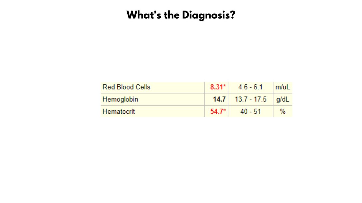
Take a look at the values in the graphic. Without looking ahead in the thread, can you make a diagnosis?
2/9
The patient has ERYTHROCYTOSIS (defined as an increased RBC count) and POLYCYTHEMIA (based on the Hct, but not the Hb).
3/9
What's up with the elevated Hct but normal Hb? Well, there is violation of the 3:1 rule which states that the Hct is normally 3x the Hb (e.g., Hct 45, Hb 15). Stated another way, the MCHC (Hb/Hct) is low, thus there is HYPOCHROMIA.
Hemophagocytic lymphohistiocytosis (HLH) is a rare, life-threatening hyperinflammatory syndrome of intense immune activation associated with fever, hepatosplenomegaly, cytopenias, hyperferritinemia, and presence of activated macrophages in hemopoietic organs.
2/6
HLH in adults is usually initiated by external causative factors such as drugs and infections or in the context of certain underlying diseases including cancer or autoimmune disease.
3/6
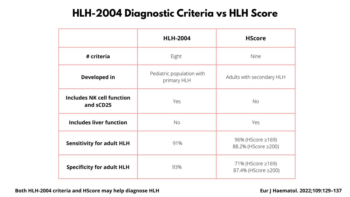
Diagnosis of HLH relies on clinical scoring systems since there no single laboratory finding has the sensitivity and specificity to definitively detect HLH. There are two commonly employed scoring systems:
I have seen a number of patients with HbSC disease lately, and thought it would be helpful to review the differences between HbSC and HbSS.
1. HbSC disease is caused by co-inheritance of the hemoglobin S (HbS) and hemoglobin C (HbC) beta globin gene mutations.
2/5
2. HbC is the second most common hemoglobin variant after HbS in the US, and third most common worldwide after HbS and HbE. HbC Confers protection against severe malaria.
3. HbSC accounts for 30% of sickle cell disease (SCD) in the US and UK.
3/5
4. Pathophysiology of HbSC disease involves:
1) RBC dehydration leading to increased HbS concentration and sickling under deoxygenated conditions.
2) HbC crystallization under oxygenated conditions.