
🧵 on our preprint with @ZaidYounes9 and team
In this study, we delve deep into COVID pathophysiology to show that:
SARS-CoV-2 infects the human bone marrow megakaryocytes, and this shapes the trajectory and severity of the illness by triggering ...
🔗biorxiv.org/content/10.110…
In this study, we delve deep into COVID pathophysiology to show that:
SARS-CoV-2 infects the human bone marrow megakaryocytes, and this shapes the trajectory and severity of the illness by triggering ...
🔗biorxiv.org/content/10.110…
... the production of pathogenic afucosylated antibodies against the spike protein.
First, some introduction in order to better understand the premise and the findings of this work:
First, some introduction in order to better understand the premise and the findings of this work:
In the last 3 years, an exhaustive amount of research has been carried out to elucidate the precise events that directly cause progression of COVID from an early mild acute illness in the first few days, to a severe/fatal form in a subset. 

Two specific early events key to such progression have been identified:
The first key event identified is the early interaction between SARS-CoV-2 and megakaryocytes (MKs) in the bone marrow.
PMID: 36920790, 35708858
The first key event identified is the early interaction between SARS-CoV-2 and megakaryocytes (MKs) in the bone marrow.
PMID: 36920790, 35708858

If MKs are infected by SARS-CoV-2, the patient is anticipated to progress to severe COVID in the ensuing days.
Vice versa, if MKs remain uninfected, the patient is expected to experience a mild course and recover.
PMID: 35708858
Vice versa, if MKs remain uninfected, the patient is expected to experience a mild course and recover.
PMID: 35708858

The other key event that is thoroughly well-established to drive severe COVID is:
The early production of a distinct form of antibody against the spike protein ("afucosylated") that delivers a double blow to the host ...
PMID: 33361116
The early production of a distinct form of antibody against the spike protein ("afucosylated") that delivers a double blow to the host ...
PMID: 33361116

This distinct anti-spike IgG antibody:
(1) Does not neutralize SARS-CoV-2
(2) And instead, triggers and directly drives the immune-mediated pathology of severe COVID
(1) Does not neutralize SARS-CoV-2
(2) And instead, triggers and directly drives the immune-mediated pathology of severe COVID
It goes without saying that upon infection with SARS-CoV-2, our immune system tries to produce antibodies against proteins of this virus, including the spike protein, with one goal:
To neutralize and clear the infection. And this happens without deviation in a majority.
To neutralize and clear the infection. And this happens without deviation in a majority.
However, not all antibodies targeting the spike protein are created equal in COVID.
Anti-spike IgGs come in two forms in this disease, with dire consequences for the patient.
Anti-spike IgGs come in two forms in this disease, with dire consequences for the patient.
Multiple studies have shown this phenomenon.
That the *quality* of antibodies produced early on in response to SARS-CoV-2 spike protein differs between individuals, and this variation determines the fate of the patient.
PMID: 33361116, 33979301, 35040666
That the *quality* of antibodies produced early on in response to SARS-CoV-2 spike protein differs between individuals, and this variation determines the fate of the patient.
PMID: 33361116, 33979301, 35040666
In fact, the detection of a distinct form of anti-spike IgG *early* in the illness accurately predicts who progresses to severe disease in the ensuing days, and who doesn’t progress and experiences only a mild illness.

So how is this variability in the anti-spike IgGs defined?
Understanding this requires a brief background on antibody structure first:
Understanding this requires a brief background on antibody structure first:
IgG antibody structure comprises of:
(1) antigen-binding region (“Fab”), where antigens such as spike protein are recognized and bound, and …
(1) antigen-binding region (“Fab”), where antigens such as spike protein are recognized and bound, and …

(2) a tail region (“Fc”) that interacts with cell surface receptors called “Fc receptors.”
These Fc receptors are found on the surface of various immune cells and platelets, which exert immune actions against the antigen recognized by the IgG.
These Fc receptors are found on the surface of various immune cells and platelets, which exert immune actions against the antigen recognized by the IgG.

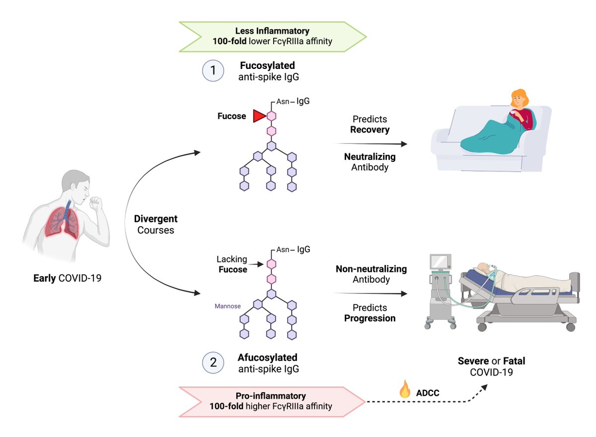
In the Fc regions of IgG, there are specific sites (at Asparagine 297) where modifications called “glycosylation” can change the inflammatory capability of each IgG.

For instance, the addition of a “fucose” moiety to this site on the Fc region would create a less inflammatory IgG.
90% of all IgGs in humans are formed this way, containing a fucose moiety, and referred to as “fucosylated” IgGs.
90% of all IgGs in humans are formed this way, containing a fucose moiety, and referred to as “fucosylated” IgGs.

On the other hand (and of relevance to COVID), the lack of fucose in the Fc region of IgG, brings about an *intensely* inflammatory quality to the antibody.
IgGs lacking fucose (“afucosylated” IgGs) have a 100-fold *higher* affinity for the FcyRIlla receptors on immune cells.
IgGs lacking fucose (“afucosylated” IgGs) have a 100-fold *higher* affinity for the FcyRIlla receptors on immune cells.

This translates into an extremely intense activation of monocytes, macrophages, NK and dendritic cells — all of which are then recruited to attack the antigen recognized and bound by the antibody.

Less than 10% of human IgGs encountered are afucosylated, as these afucosylated IgGs are intensely inflammatory and can cause severe immune-mediated pathology.
Our immune system relies on fucosylated IgGs — with much lower inflammatory quality than the afucosylated ones — to respond to a vast majority of infectious pathogens.

However, afucosylated responses are found in response to specific situations.
Infections with enveloped viruses such as SARS-CoV-2, dengue virus, and HIV can be associated with afucosylated IgG production against viral antigens.
Infections with enveloped viruses such as SARS-CoV-2, dengue virus, and HIV can be associated with afucosylated IgG production against viral antigens.
Afucosylated IgG responses are also seen in reaction to foreign antigens on blood cells such as platelets.
PMID: 24243971
PMID: 24243971

In the case of COVID, multiple prominent studies have demonstrated the detection of afucosylated IgGs against the spike protein in severe COVID.
PMID: 33361116, 33979301
PMID: 33361116, 33979301

These studies have also shown that not only afucosylated anti-spike IgGs are present in severe COVID, but also …
their presence is an early predictor and a direct driver of progression to severe COVID.
their presence is an early predictor and a direct driver of progression to severe COVID.
In two prospective cohorts of outpatients with early COVID who underwent blood draws ...
the detection of these afucosylated IgGs against the spike protein was shown to determine the fate of these individuals later on.
the detection of these afucosylated IgGs against the spike protein was shown to determine the fate of these individuals later on.
Those with an early production of afucosylated anti-spike IgG detected on, for instance day 5 of illness as outpatient, reliably progressed to severe COVID on subsequent days, whereas …
On the other hand, the patients ...
On the other hand, the patients ...

... who only formed normally fucosylated IgGs against the spike protein experienced only a mild course and recovered.

The timing of antibody formation mattered too.
In those who progressed to severe disease, the afucosylated (and non-neutralizing) IgGs were formed prior to the normally fucosylated (and neutralizing) IgGs against the spike protein.
In those who progressed to severe disease, the afucosylated (and non-neutralizing) IgGs were formed prior to the normally fucosylated (and neutralizing) IgGs against the spike protein.

Something … somehow … pushed these unfortunate individuals to form a non-neutralizing and pathogenic afucosylated response against the spike protein …
And these pathogenic antibodies were formed much earlier than the neutralizing ones that clear the virus.
And these pathogenic antibodies were formed much earlier than the neutralizing ones that clear the virus.
Mechanistically, these studies also clearly show how the formation of these afucosylated anti-spike IgGs are key drivers of the immune-mediated pathology in severe COVID …
Recall that lack of fucose in the anti-spike IgGs triggers an intense activation of monocytes, macrophages, NK and dendritic cells — all targeted any and all tissue harboring the spike protein
In other words, these afucosylated IgGs order our own immune system to most ferociously attack and eliminate any trace of spike protein in the body.

The only problem becomes that …
the spike protein is broadly expressed on the cell surface of many infected human tissues during COVID.
And our own body becomes the collateral damage to our immune system’s reaction by way of these afucosylated IgGs
the spike protein is broadly expressed on the cell surface of many infected human tissues during COVID.
And our own body becomes the collateral damage to our immune system’s reaction by way of these afucosylated IgGs
This is why in severe COVID, most successful therapeutics are immune-modulators such as steroids, JAK/STAT inhibitors, anti-TNFs, etc.
These afucosylated responses are *not* unique to severe COVID. We’ve seen them before.
They also drive severe dengue.
PMID: 31765380, 34083490
They also drive severe dengue.
PMID: 31765380, 34083490

Dengue virus also happens to another enveloped RNA virus like SARS-CoV-2, that also infects bone marrow megakaryocytes like SARS-CoV-2.

In addition to these, afucosylated responses are also seen in HIV, but this time, they act in a beneficial fashion by conferring improved viral suppression.
HIV is yet another enveloped virus that not only infects MKs, but also ...
HIV is yet another enveloped virus that not only infects MKs, but also ...
... uses MK infection as a reservoir to cause persistence despite patients taking highly-active antiretroviral therapy.
PMID: 32188724
PMID: 32188724

We thought there may be a pattern here, perhaps.
And naturally, this is where shaping of our hypothesis began.
Is there a relationship between enveloped viruses infecting MKs, and this infection causing afucosylated responses toward antigens of these enveloped viruses?
And naturally, this is where shaping of our hypothesis began.
Is there a relationship between enveloped viruses infecting MKs, and this infection causing afucosylated responses toward antigens of these enveloped viruses?

What do we know about the contexts and antigens that are associated with afucosylated responses?
Owing to a comprehensive study by Larsen et al. (PMID 33361116), we know that ...
Owing to a comprehensive study by Larsen et al. (PMID 33361116), we know that ...

Afucosylated responses can occur in response to a limited set of stimuli including …
(1) Enveloped virus antigens
(2) Alloantigens on surface of platelets
(3) Alloantigens on surface of RBCs
(1) Enveloped virus antigens
(2) Alloantigens on surface of platelets
(3) Alloantigens on surface of RBCs

In contrast (bottom row):
(1) Soluble proteins
(2) Bacterials antigens, and
(3) Non-enveloped virus antigens
are not known to cause afucosylated responses — and instead form the typical fucosylated IgGs only.
(1) Soluble proteins
(2) Bacterials antigens, and
(3) Non-enveloped virus antigens
are not known to cause afucosylated responses — and instead form the typical fucosylated IgGs only.

To dig deeper …
In the case of enveloped viruses, not all viral proteins are known to trigger afucosylated responses.
Larsen et al. have demonstrated that the location of the viral protein matters.
Surface expression is the differentiator.
In the case of enveloped viruses, not all viral proteins are known to trigger afucosylated responses.
Larsen et al. have demonstrated that the location of the viral protein matters.
Surface expression is the differentiator.

Surface proteins of enveloped viruses can cause afucosylated responses. An example of a surface protein would be the spike protein of SARS-CoV-2.
In contrast, viral proteins without any surface expression do not induce afucosylated responses.
In contrast, viral proteins without any surface expression do not induce afucosylated responses.

Larsen et al. showed in the case of severe COVID patients, afucosylated IgGs are only detectable against the spike protein (has surface expression) …
Whereas very little to no afucosylated IgGs are formed against the nucleocapsid protein (no surface expression).
Whereas very little to no afucosylated IgGs are formed against the nucleocapsid protein (no surface expression).

We also know that afucosylated responses are seen in response to foreign antigens expressed on the surface of platelets.
This is best illustrated in the case of a fetal platelet disorder called FNAIT.
PMID: 24243971
This is best illustrated in the case of a fetal platelet disorder called FNAIT.
PMID: 24243971

Further refining our hypothesis based on these lines of evidence …
We began to wonder whether ...
SARS-CoV-2 can infect platelets and pathogenically embed its spike protein on the surface of platelets ➡️leading to an afucosylated response and the associated immunopathology.
We began to wonder whether ...
SARS-CoV-2 can infect platelets and pathogenically embed its spike protein on the surface of platelets ➡️leading to an afucosylated response and the associated immunopathology.

But we hit a roadblock there.
Several prominent studies clearly show that platelets are infected by SARS-CoV-2 during COVID.
None, however, were able to visualize evidence of spike protein expression on the surface of platelets.
Several prominent studies clearly show that platelets are infected by SARS-CoV-2 during COVID.
None, however, were able to visualize evidence of spike protein expression on the surface of platelets.
I even pursued personal communication with the authors of these studies over the last 2 years, who confirmed that …
... although the spike protein was found in the cytoplasm of infected platelets, and such finding was highly predictive of severe disease progression, no evidence of spike protein expression was found on the surface of platelet membrane.
So, our attention switched to platelet progenitors, megakaryocytes, to explore our suspicion about the spike protein expression on the surface of these cells.
Multiple human studies thus far have established that SARS-CoV-2 infects MKs in the bone marrow (all in postmortem samples of bone marrow)
Two studies in particular demonstrated that not only SARS-CoV-2 infects MKs, but also …
The viral infection of bone marrow MKs — similar to the case of afucosylated anti-spike IgGs — was an early predictor of progression to severe COVID.
PMID: 36920790, 35708858
The viral infection of bone marrow MKs — similar to the case of afucosylated anti-spike IgGs — was an early predictor of progression to severe COVID.
PMID: 36920790, 35708858

Strikingly, virus-infected MKs were shown to transfer viral antigens including the spike protein (red) to emerging platelets during differentiation to proplatelets and platelets (Fortmann et al.)

What was not explored in this study was whether the spike protein is specifically expressed on the surface of resultant platelet progenitors emerging from the virus-infected MKs.
Such finding — similar to platelet alloantigens encountered in FNAIT — would provide a plausible pathway by which afucosylated IgGs form against the spike protein.

Moreover, thus far no group has explored the possibility of whether virus-infected bone marrow MKs may be the key trigger for the production of the pathogenic afucosylated IgGs against the spike protein.

These two questions formed the basis of our study.
The schematic below summarizes the four parts of our hypothesis.
The schematic below summarizes the four parts of our hypothesis.

Based on the described hypotheses, experiment was designed as depicted in this diagram.

After obtaining IRB and approval from ethics committee, human bone marrow megakaryocytes were isolated from hospitalized subjects who were in the early days of experiencing severe COVID.

Subsequently, human bone marrow MKs were isolated from the bone marrow aspirate.
PCR confirmed the infection of bone marrow MKs by SARS-CoV-2 in these living donors in early stage of severe COVID.
PCR confirmed the infection of bone marrow MKs by SARS-CoV-2 in these living donors in early stage of severe COVID.

Next …
Virus-infected MKs were grown and allowed to differentiate under favorable conditions and assessed for evidence of spike protein expression on the surface of resultant platelet progenitors.
Virus-infected MKs were grown and allowed to differentiate under favorable conditions and assessed for evidence of spike protein expression on the surface of resultant platelet progenitors.

Proplatelets emerging from the virus-infected MKs were labeled to identify their periphery (α-tubulin, green) and to identify any evidence of spike protein (red).
Indeed, we found the spike protein expressed on the surface of virus-infected proplatelets!
Indeed, we found the spike protein expressed on the surface of virus-infected proplatelets!

We then pursued our second question.
Virus-infected human bone marrow MKs were injected intravenously to transgenic mice harboring human ACE2 to see whether it triggers the formation of fucosylated IgGs against the spike protein.
Virus-infected human bone marrow MKs were injected intravenously to transgenic mice harboring human ACE2 to see whether it triggers the formation of fucosylated IgGs against the spike protein.

First:
We found that injection of virus-infected human MKs into transgenic mice significantly promoted the formation afucosylated IgG antibodies against the spike protein
To our knowledge, this is the 1st instance of afucosylated IgGs detection against a viral antigen in vivo.
We found that injection of virus-infected human MKs into transgenic mice significantly promoted the formation afucosylated IgG antibodies against the spike protein
To our knowledge, this is the 1st instance of afucosylated IgGs detection against a viral antigen in vivo.

Second:
The mortality rate in the infected mice was proportional to the level of afucosylated anti-spike IgG produced.
The mortality rate in the infected mice was proportional to the level of afucosylated anti-spike IgG produced.

This finding suggests that the afucosylated anti-spike IgG antibodies were pathogenic and contributed significantly to the severity of COVID-19 in mice - as is the case in humans.

Third:
The plasma levels of PF4 and serotonin, as soluble markers of platelet activation and/or granule release, were significantly higher in the infected mice compared with the uninfected mice.
The plasma levels of PF4 and serotonin, as soluble markers of platelet activation and/or granule release, were significantly higher in the infected mice compared with the uninfected mice.

Lastly, and perhaps most importantly:
Injection of virus-infected MKs into the circulation of these mice (without inhalation of the virus) mimicked the lung pathology seen in human cases of severe COVID:
Injection of virus-infected MKs into the circulation of these mice (without inhalation of the virus) mimicked the lung pathology seen in human cases of severe COVID:

Inflammatory cell infiltrate in the lung, followed by clot formation in the pulmonary vascular lumen, and distortion of the lung tissue.

In summary, to the best of our knowledge, this is the first study that demonstrates direct evidence that bone marrow megakaryocytes are infected early on by SARS-CoV-2 in those experiencing severe COVID.

Additionally, to the best of our knowledge, this is the first study that demonstrates proplatelets express SARS-CoV-2 spike protein on the surface of their membranes during differentiation from virus-infected human MKs.
This surface expression of spike protein matters …
This surface expression of spike protein matters …

... as it likely explains the context within which our immune system decides to switch gears and form highly inflammatory afucosylated antibodies against the spike protein.
In millions infected by this virus, the immune response follows a predictable pattern.
Virus infects the respiratory tract. Immune system reacts in a typical fashion, and forms neutralizing fucosylated antibodies against the spike protein.
Virus clears. Acute disease resolves.
Virus infects the respiratory tract. Immune system reacts in a typical fashion, and forms neutralizing fucosylated antibodies against the spike protein.
Virus clears. Acute disease resolves.

In a subset who progress to severe COVID, the antibody response is atypical — and actually causes disease progression.
Our proposed mechanism is that in these instances, the virus likely breaches the respiratory tract, reaches the bloodstream, and infects the bone marrow MKs.
Our proposed mechanism is that in these instances, the virus likely breaches the respiratory tract, reaches the bloodstream, and infects the bone marrow MKs.
As the virus infects MKs, and expresses the spike protein on the surface of emerging proplatelets, the immune system senses a much more 🔥dangerous situation.
Body’s own circulating blood cells are now infected — beyond the respiratory tract, beyond the oropharynx.
Body’s own circulating blood cells are now infected — beyond the respiratory tract, beyond the oropharynx.
The infection of circulating blood cells is likely a highly alarming 🚨context to the immune system.
To combat viral spread now aided by far-reaching infected platelets, a most potent inflammatory reaction is called into action by creating afucosylated IgGs against spike protein
To combat viral spread now aided by far-reaching infected platelets, a most potent inflammatory reaction is called into action by creating afucosylated IgGs against spike protein
This leads to an intense FcyRIlla-mediated activation of monocytes, macrophages, NK and dendritic cells systemically, attacking every tissue harboring spike protein.
That is, unfortunately, a large part of body's tissues, including the vasculature, lung, neural tissue, etc.
That is, unfortunately, a large part of body's tissues, including the vasculature, lung, neural tissue, etc.

... causing widespread immuno-thrombosis, and immune-mediated injury to vasculature, lungs, blood-brain barrier, nervous system, and other tissues in the body.

In summary, we provide preliminary evidence that SARS-CoV-2 infects human bone marrow megakaryocytes, and this infection of megakaryocytes is directly responsible for triggering the immune-mediated pathology of severe COVID.
biorxiv.org/content/10.110…
biorxiv.org/content/10.110…
We acknowledge that this is a preprint, and has not been peer-reviewed.
We also acknowledge that we have a significant amount of future work left to be done to better characterize the preliminary findings described in this work.
We also acknowledge that we have a significant amount of future work left to be done to better characterize the preliminary findings described in this work.

If your group has expertise in glycobiology, please consider reaching out to us as we are open to collaboration to decipher the detailed mechanisms of the findings in this work.
We appreciate your patience in reading through this long thread. And we encourage you to read our preprint below for further details.
biorxiv.org/content/10.110…
biorxiv.org/content/10.110…
Lastly, we would like to thank the entire team of Dr. Younes Zaid @ZaidYounes9, and acknowledge the generous funding by Balvi and @VitalikButerin to allow us to continue our work.

• • •
Missing some Tweet in this thread? You can try to
force a refresh


