Here is analysis of HA mutations in H5 influenza case in Missouri resident without known contact w animals or raw milk.
TLDR: there is one HA mutation that strongly affects antigenicity, and another that merits some further study.
TLDR: there is one HA mutation that strongly affects antigenicity, and another that merits some further study.
As background, CDC recently released partial sequence of A/Missouri/121/2024, which is virus from person in Missouri who was infected with H5 influenza.
Here I am analyzing HA protein from this release, GISAID accession EPI_ISL_19413343cdc.gov/bird-flu/spotl…
Here I am analyzing HA protein from this release, GISAID accession EPI_ISL_19413343cdc.gov/bird-flu/spotl…
Sequence covers all of HA except signal peptide, and residues 325-351 (sequential numbering) / 312-335 (H3 numbering). The missing residues encompass HA1-HA2 boundary, and any missed mutations there unlikely to affect antigenicity or receptor binding, but could affect stability.
In sequenced part of HA, are 4 mutations relative to current H5 2.3.4.4b candidate vaccine virus, 2 of which are also mutations relative to most (but not all) cattle HAs
The 2 mutations in all cattle viruses unlikely to be relevant, so I will focus on the 2 more unique mutations
The 2 mutations in all cattle viruses unlikely to be relevant, so I will focus on the 2 more unique mutations
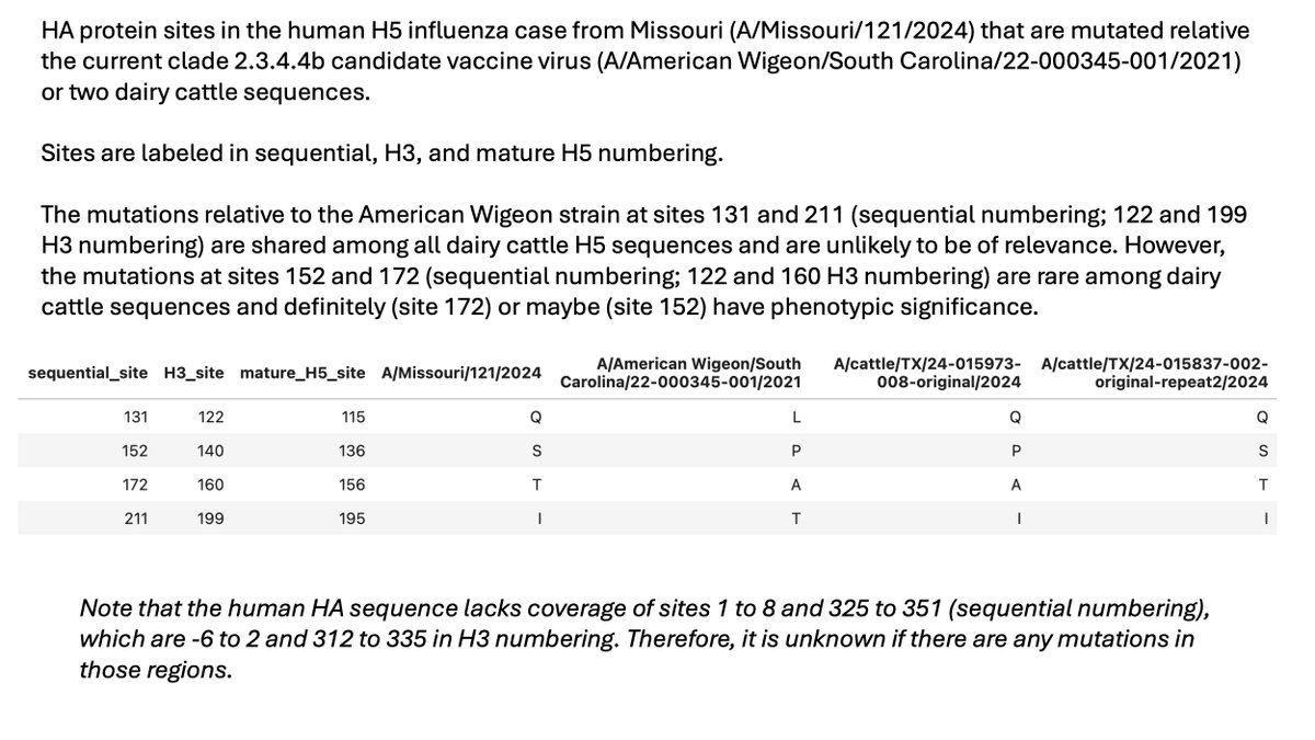
First mutation is A160T (H3 numbering), which is A172T in sequential numbering and A156T in mature H5 numbering.
From our deep mutational scanning (), we know this mutation strongly reduces neutralization by sera from current candidate vaccine virus. biorxiv.org/content/10.110…
From our deep mutational scanning (), we know this mutation strongly reduces neutralization by sera from current candidate vaccine virus. biorxiv.org/content/10.110…

Because deep mutational scanning flagged this mutation, @bdadonaite previously performed neutralization assays showing A160T (a la A172T, A156T) causes a 10- to 100-fold drop in neutralization by sera from ferrets exposed to the current candidate vaccine virus.

The A160T (a la A172T, A156T) mutation is found in a handful of dairy cattle H5 sequences, although most lack the mutation. This can be seen by looking at @LouiseHMoncla’s nextstrain build: nextstrain.org/avian-flu/h5n1…
This mutation underscores challenges of candidate vaccine virus approach
We don’t know what (if any) virus might eventually spread in humans, but would be unfortunate to stockpile American Wigeon or Astrakhan based vaccine, only to have A160T virus spread
We don’t know what (if any) virus might eventually spread in humans, but would be unfortunate to stockpile American Wigeon or Astrakhan based vaccine, only to have A160T virus spread
https://x.com/jbloom_lab/status/1819746067317195184
Fortunately, CDC seems attentive to this issue, as their report correctly () notes the potential impact of this mutation on candidate vaccine viruses.cdc.gov/bird-flu/spotl…
Second mutation is P140S (H3 numbering), which is P152S in sequential numbering and P136S in mature H5 numbering.
This mutation is only found in a single sequenced dairy cattle virus, which has an HA identical to the Missouri case over the covered region.
This mutation is only found in a single sequenced dairy cattle virus, which has an HA identical to the Missouri case over the covered region.

Although P140S is on head of HA in classically defined antigenic region A, deep mutational scanning shows it does not have much effect on neutralization by ferret or mouse sera elicited by candidate vaccine viruses (). dms-vep.org/Flu_H5_America…

However, some attention should be given to possibility P140S (a la P152S, P136S) could slightly impact receptor binding. In our deep mutational scanning (), P140S modestly improves entry into a2-6 expressing cells.dms-vep.org/Flu_H5_America…
I emphasize any impact of P140S is speculative: in deep mutational scanning its effect is modest, and we have not validated that initial finding further. Additionally, P140S is not a receptor contact site, although it is proximal to receptor binding loops.
However, further study of P140S (a la P152S, P136S) is merited by combined facts that
(1) it causes modest improvement a2-6 usage in deep mutational scanning, and
(2) it was found in human Missouri case despite being in only one of many known dairy cattle sequences.
(1) it causes modest improvement a2-6 usage in deep mutational scanning, and
(2) it was found in human Missouri case despite being in only one of many known dairy cattle sequences.
Thanks to CDC (and Missouri Dept of Health) for releasing the partial sequences alongside their careful analysis ()cdc.gov/bird-flu/spotl…
Deep mutational scanning data I used to interpret the mutations is all available at dms-vep.org/Flu_H5_America…
• • •
Missing some Tweet in this thread? You can try to
force a refresh