MariaGutschi✝️📿💊👵🦠 Profile picture
Dec 1 10 tweets 3 min read Read on X
1/ 🧵Pfizer’s new mRNA flu trial quietly revealed the problem they can’t quite admit....

It’s not the spike. It’s the platform. It's the LNPs.
What did the data show?
nejm.org/doi/full/10.10…
2/Pfizer’s mRNA flu vaccine = same tech as COVID shots, just a new mRNA coding for the FLU antigen.
So what happened?
Strong fevers, swollen nodes, fatigue, pain which was identical to the COVID pattern.
Same signature, but NO SPIKE PROTEIN.
Does that mean the toxic driver is the lipid nanoparticle (LNP) system itself?
3/ Even worse: in the >65 year-old trial dataset (quietly posted on , no press release)…
There’s an important renal (kidney) signal.
Serious kidney adverse events in healthy seniors who were screened to exclude any chronic issues including kidney impairment.
That’s a major pharmacologic red flag.clinicaltrials.gov
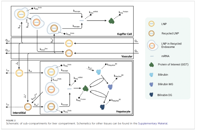
4/ LNPs are not “fat bubbles.”
They’re ionizable cationic amphiphiles (CADs) which are positively charged at acidic pH inside the endosomes.
These molecules are biologically active: they fuse with cell membranes, get trapped in lysosomes, and can trigger phospholipidosis and mitochondrial stress.
They behave like drug molecules, not inert carriers.
5/ They're like mini‑detergents or steel wool scrubbing your cell membranes (not quite, but well, you get the picture).
They dissolve INTO cell membranes, scramble lipids, activate complement cascades ( fevers/chills), and distribute through liver, spleen, kidney, ovaries, etc
6/ If the spike is removed but the inflammation remains, you’ve identified the real culprit.
The antigen (spike protein, hemagluttinin, the RSV proteint) isn’t driving these side effects.
The "platform" is.

Pfizer’s own flu trial data quietly confirmed it.
7/ these kidney signals, may be associated with the LNPs CAD-like activity, similar to how aminoglycosides, another CAD causes kidney damage (i.e. gentimicin, tobramycin, amikacin etc)

Other possibilities include microclots gumming up the renal tubules but this too would need to be initiated by the LNPs because there is no spike protein in these mRNA influenza vaccines..
8/ I think regulators should look at standard CAD pharmacology screens for the LNPs:
• phospholipidosis assays
• lysosomal trapping quantification
• kidney & liver ultrastructure studies
• full lipid biodistribution & metabolite tracking
These are basic toxicology 101
9/ The LNPs are not inert. They’re active compounds.
They act like amphiphilic drugs, pharmacological detergents, which cause inflammation, cellular stress, and a kidney signal that cannot be ignored imho.

Should amphiphilic ionizable lipids, be used for annual or chronic injections? The answer is obvious.
10/ When the plug (antigen) changes but the reaction stays the same, the platform IS the drug.
And the drug is toxic. We need the FDA/EMA/HC to act
Read my full analysis here: mariagutschi.substack.com/p/what-the-new…

• • •

Missing some Tweet in this thread? You can try to force a refresh
 

Keep Current with MariaGutschi✝️📿💊👵🦠

MariaGutschi✝️📿💊👵🦠 Profile picture

Stay in touch and get notified when new unrolls are available from this author!

Read all threads

This Thread may be Removed Anytime!

PDF

Twitter may remove this content at anytime! Save it as PDF for later use!

Try unrolling a thread yourself!

how to unroll video
  1. Follow @ThreadReaderApp to mention us!

  2. From a Twitter thread mention us with a keyword "unroll"
@threadreaderapp unroll

Practice here first or read more on our help page!

More from @CanningPharm

Nov 27
1/ So this is how the vaccine "works" as per this paper. Can you count the lies/misinformation? Image
2/ There are really 2 models in this paper but I will focus mostly on the first one.
Model 1:
Visual and written depiction of how mRNA vaccines work by 1. mRNA enters cytosol, 2. ribosomes produce spike antigen, 3. immune memory results.

Model 2: Depiction of how cells protect themselves from foreign DNA, that is the nucleus has a membrane barrier and “a tiny fragment of foreign DNA is harmless because it lacks the tools to insert itself.”
3/There is no discussion of:

The chemical structure or bio-distribution of lipid nanoparticles (LNPs). In fact LNPs aren't mentioned at all.

The presence or biological activity of residual plasmid DNA or other manufacturing impurities which have biological activity.

Ionizable lipid persistence, organ accumulation, or its inflammatory properties.

Immune activation through PPRA or lysosomal disturbance.

They take as axiomatic that: “mRNA vaccines do not enter the nucleus where DNA resides and cannot change or influence our genes.” What happens when the nuclear envelope dissolves during mitosis.

They pass over actual kinetic, biochemical, and toxicological complexity of the delivery vehicle.
Read 4 tweets
Nov 25
1/10 🧵I am in rant mode today. I feel invisible. I am often dismissed as a "pill counter" despite knowing formulation, stability, & drug kinetics. And it is so important re mRNA-LNP platform. Thread on why it matters, and how AI is making it WORSE. #mRNA
Tweet 2/10:
40 yrs as a pharmacist (and only 2 behind a counter). I've crunched LNP biophysics & manufacturing deviations for 4 yrs. But once I reveal my background? Interviews vanish. Academia, biotech, regulators: often treat us as secondary voices. It's structural, not personal. 💔
Full disclosure: I am a hospital and highly trained pharmacist, clinician educator, wrote chapters in textbooks, designed award winning programs, worked at a national level, so admittedly not your average pharmacist.
Tweet 3/10:
Enter AI. Asked ChatGPT about pharmacist Christine Delpetre's claims on mRNA batch variability, DNA contamination, & EMA leaks. Solid data, yet it downplays her cuz she's "just a pharmacist." Same facts from a virologist? "Authoritative." Holy Toledo! 🤯Image
Image
Read 10 tweets
Sep 9, 2024
1/ Here is the standard PBPK model they used. First look at where and how much blood flow. They used IV administration. panel A
Image
2/ so the organ exposed the most is the lung, kidneys, GI tract to liver based on blood flow. But we hardly hear about transfections in kidney or lung. There is a disconnect between uptake into tissues (min) and expression of protein (hrs).
3/ for now, just look at the curves of the red lines which is distribution of the LNPs which are labelled with iodine. This is tissue uptake or transfection. Image
Read 8 tweets
Apr 20, 2024
1/The pharmacokinetics of LNPs are exceedingly complex but this paper is the best so far in trying to apply traditional physiologic-based pharmacokinetic modelling with some empirical validation. Let's see if I can explain it to the best of my ability (note paper at end of thread)Image
2/That figure is a schematic of a liver Kupffer cell, blood or vascular flow, a hepatocyte and interstitial fluid.
through the liver
2. LNPs passively infiltrate the interstitial space and can either be taken up by the Kupffer cells or hepatocyte (transfection)
3. or flow back into lymph nodes and then back again into the vasculature and are recycled
4. once the LNP transfects either the Kupffer or hepatocytes they can undergo one of 4 processes
3/
a. degradation through the endosomes and then lysosomes (can take a long time) and are stuck there
b. mRNA escape into the cytoplasm (the desired effect), then production and degradation of the expressed protein
c. transition into recycled endosomes and exocytosed
d. degradation of escaped mRNA in the cell

These exocytosed LNPs likely do not have the same surface properties of the original LNPs but can still travel and transfect other cells, and are the cause of "shedding" imho.

Also, the recycled but plain LNPs which travel through the vascular and lymph circulation produce a second peak in LNP kinetics sometimes seen experimentally
Read 7 tweets
Aug 6, 2023
1/The mRNA in these vaccines is not immunogenic and thus cannot function as a built-in adjuvant. Instead, it seems that the delivery of the LNP stimulates the innate immune system and functions as an adjuvant. Or is it the only adjuvant?

nature.com/articles/s4159…
2/But none of the innate immune sensors, including TLR2, TLR3, TLR4, TLR5, TLR7 and cGAS, seemed to be needed for the immunogenicity of this vaccine.

Only MDA-5, a receptor for long double-stranded RNA, was shown to be important for type I interferon responses and as a built-in adjuvant pathway for antigen-specific CD8+ T cell responses.

There’s dsRNA in those vials as a contaminant.
3/Oh dsRNA as an adjuvant? A pretty good one in fact.

Synthetic dsRNA analogues have been tested as vaccine adjuvant against viral infections in animal models.

But not so fast.

However, a dsRNA receptor, TLR3 can be expressed in tumor cells while other members of TLR family, for example, TLR4 and TLR2 have been shown to promote tumor progression, metastasis, and chemoresistance

Ooops

ncbi.nlm.nih.gov/pmc/articles/P…
Read 9 tweets
Nov 23, 2022
Well now I have a better idea why autophagy and intermittent fasting works to get rid of the spike protein.
ncbi.nlm.nih.gov/pmc/articles/P…
So Moderna looked for ways to make its proteins last longer in the body, including de-ubiquitination. 😲 Image
They did homology searches to identify related sequences from other species. Found amino acids that could be changed to stabilize folding and remove ubiquitination sites. They did this for phenylalanine hydroxylase. Bet they did this for the SP as well. Image
Read 6 tweets

Did Thread Reader help you today?

Support us! We are indie developers!


This site is made by just two indie developers on a laptop doing marketing, support and development! Read more about the story.

Become a Premium Member ($3/month or $30/year) and get exclusive features!

Become Premium

Don't want to be a Premium member but still want to support us?

Make a small donation by buying us coffee ($5) or help with server cost ($10)

Donate via Paypal

Or Donate anonymously using crypto!

Ethereum

0xfe58350B80634f60Fa6Dc149a72b4DFbc17D341E copy

Bitcoin

3ATGMxNzCUFzxpMCHL5sWSt4DVtS8UqXpi copy

Thank you for your support!

Follow Us!

:(