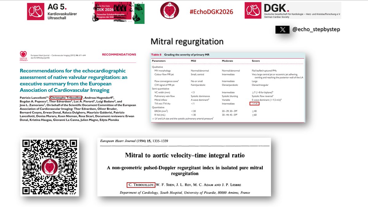
Current #guidelines recommend the mitral inflow/LVOT VTI ratio as an easy-to-measure semiquantitative index for isolated primary MR (>1.4 severe, <1 mild). But how robust is the evidence behind it? Let's take a closer look at this widely cited parameter. #EchoFirst Part 1 

The mitral inflow/LVOT VTI ratio was first introduced as a semiquantitative parameter for grading primary mitral regurgitation in the 2013 EACVI #recommendations. This recommendation was based on the original study by Christophe Tribouilloy et al., published in 1994. Part 2
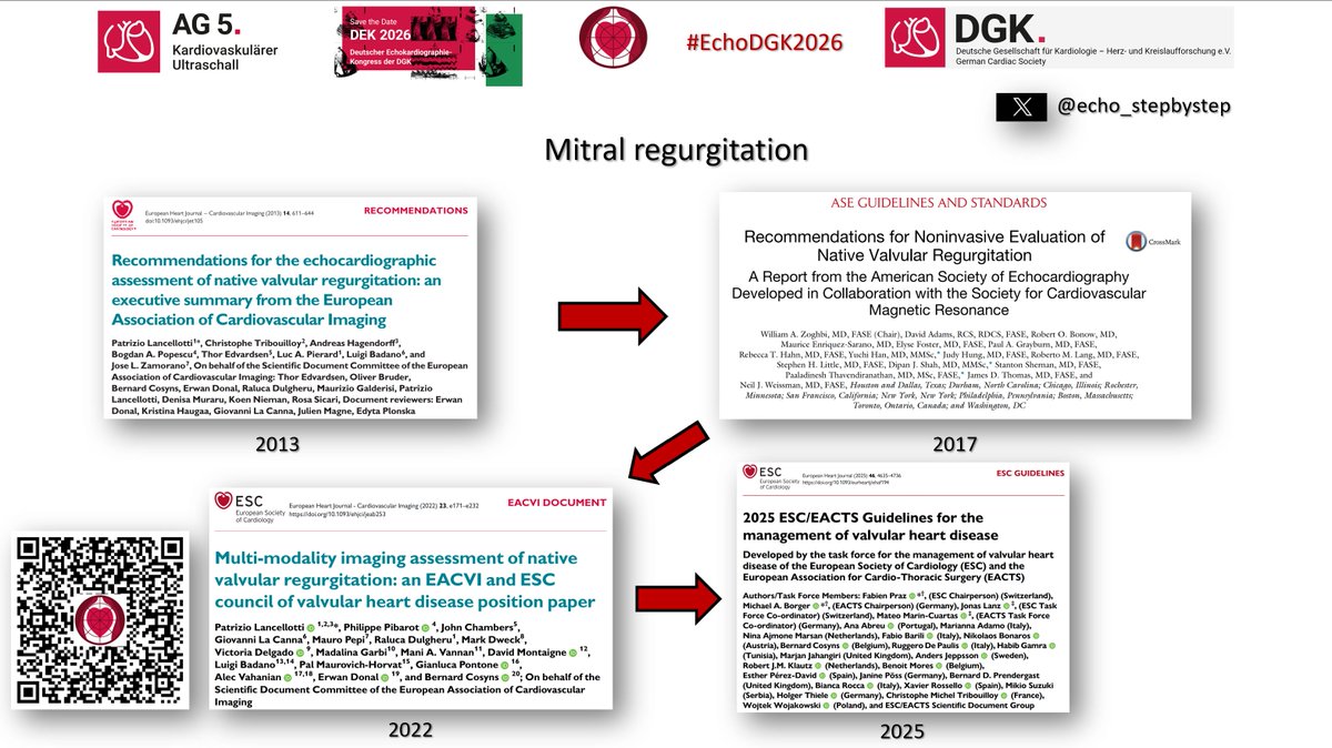
4 years later, the same parameter was adopted in the 2017 ASE recommendations under the leadership of @WilliamZoghbi, again citing the original 1994 study by C. Tribouilloy. It was subsequently retained in the 2022 EACVI/ESC Council Position Paper @ the 2025 ESC VHD Guidelines.
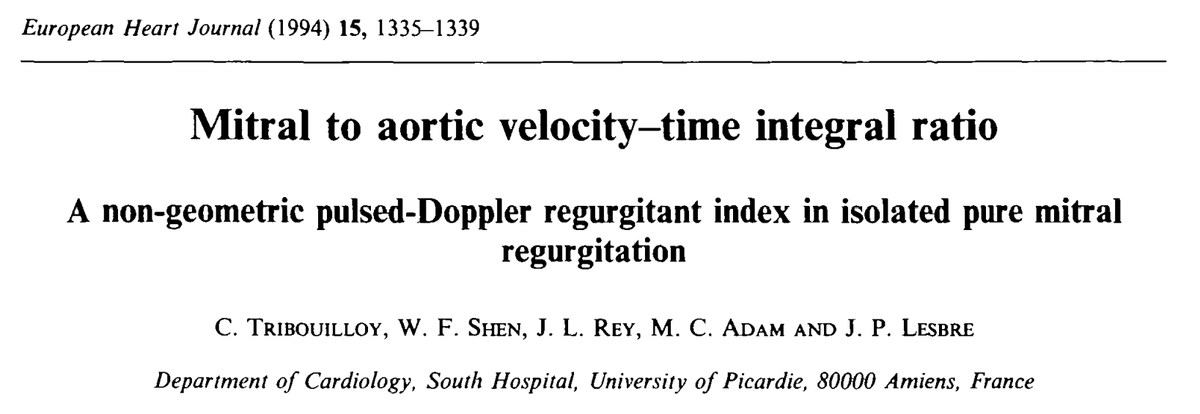
@WilliamZoghbi Let's take a closer look at the original study by Christophe Tribouilloy et al. (1994) and critically examine the evidence that has supported this recommendation for more than three decades. Part 4.
@WilliamZoghbi In the original study, mitral inflow VTI was measured at the mitral leaflet tips in the apical 4-chamber view, while aortic VTI was obtained at the aortic annulus in the apical 5-chamber view. The MV/LVOT VTI ratio was calculated from the average of three measurements. Part 5
@WilliamZoghbi But was this Doppler acquisition strategy appropriate? Comparing PW Doppler measurements of mitral inflow (A4C vs. APLAX) and LVOT flow (A5C vs. APLAX) reveals differences of up to 14%. Part 6

@WilliamZoghbi There is, however, a comparable imaging plane that yields LVOT VTI values equivalent to those obtained from the APLAX view: the modified anteriorly tilted two-chamber view. This view may provide a more consistent approach for acquiring LVOT Doppler measurements. Part 7
@WilliamZoghbi The modified anteriorly tilted two-chamber view is generated by cranial angulation of the transducer about its long edge while maintaining the apical acoustic window. Part 8
@WilliamZoghbi In A4C, transmitral inflow is often insonated at a larger angle to the ultrasound beam. In APLAX, flow is nearly parallel to the beam, potentially reducing angle-dependent error and resulting in higher E-wave velocities and more accurate PW Doppler measurements. Part 9
• • •
Missing some Tweet in this thread? You can try to
force a refresh