
Our 2nd & final thread in a review of key findings of pathologist Prof. Arne Burkhardt, a notable pioneer in the histological investigation of tissue/cell damage from the m-RNA injections. English subtitled video(s) and a transcript can be found here: docs.shortxxvids.com/burkhardt_anal…
The first thread, for anyone who missed it, can be found here. Please note that since this thread, all translation questions were kindly resolved by Prof. Burkhardt himself & the text has been supplemented with additional definitions of medical terms.
These threads were created in collaboration with Saji Hameed, professor for Climate Science at the Aizu University in Fukushima & I want to acknowledge his great contributions and insights brought to presenting key aspects of Burkhardt's findings.
<u-aizu.ac.jp/research/facul…>
Prof. Burkhardt packed much data into a one hour presentation. However, with such a time constraint, the discussion is necessarily hurried and in parts lacks sufficient detail. Therefore, we will focus on a few observations which are adequately discussed.
To orient the curious reader, we provide a brief and focused introduction to histological analysis. Our discussion is of necessity simplistic and general. If you want to learn more, a good starting place is histology.siu.edu/intro/tissprep…
Histological analysis uses stains to artificially color various parts of cells and tissues. A commonly used stain is H&E. Here, H refers to Hematoxylin, a purple dye which selectively stains the nuclei of cells. E refers to Eosin, a red dye which stains proteins in our organs.
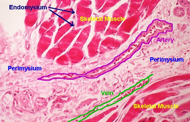
Look at a section of muscle stained by H&E from this page. pathguy.com/histo/063.html
The areas in red contain bundles of muscle fibers. At the edge of the red areas, the barely visible (you need to look hard to see these) elongated blue or purple areas are the nuclei of cells called myocytes. These cells synthesize the fibers that make up the muscle.
The pinkish areas in the same slide can in very general terms be called the Extra Cellular Matrix (ECM). It consists of polysaccharides (sugar polymers) and protein fibers such as collagen and elastin.
Here, the ECM not only surrounds the groups of muscle fibers, but also each individual fiber. The curious may find more information here biopathica.co.uk/documents/stud…
By the way, the figure also shows a vein and a nerve embedded inside regions of ECM. If you cannot see them, please see the labeled version of this slide here pathguy.com/histo/063z.jpg
One of the key findings by Prof. Burkhardt is the damage to elastic fibers and muscles of the blood vessels. To understand his discussion about vascular damages, we need to know how blood vessels look under normal and abnormal conditions.
This picture shows a collection of blood vessels and nerves artificially colored using a stain called elastin histology.siu.edu/crr/CR025b.htm
This stain provides more information about the different kind of protein fibers in a sample.
See or ncbi.nlm.nih.gov/books/NBK51905… if you would like to know more about elastin stains.
3 distinct layers surround the artery shown here: histology.siu.edu/crr/CR023b.htm The tunica media is the thick purple layer with visible nuclei, those of myocytes. It is sandwiched by the tunica intima (deep purple) & tunica adventitia (pale pink). These 3 layers form the arterial wall.
One important point is that the thickness of the arterial wall is dependent on the size of the artery. Larger arteries have thicker walls and the wall gets thinner as the artery branches and become smaller. An artery that develops a disproportionately thick wall signals trouble.
Toxins such as the chemotherapy drug Bleomycin can damage arteries.
ncbi.nlm.nih.gov/pmc/articles/P…
In the figure, the column in the middle shows an abnormally thick artery wall in a mouse injected with this drug.
The three rows show the same artery artificially colored using 3 different stains. The damaged artery (left column) may be compared to the normal artery in the rightmost column. The abnormal thickening is associated with damage to the Extra Cellular Matrix.
Burkhardt's main findings in large blood vessels are alarming — many cases of texture changes to the aorta wall. In 40 cases, these changes developed into aortal 'dissections' = tearing apart of the vessel wall layers. In 5 cases, the dissections developed into perforations.
In the past, such textural changes to vessel walls were known to the medical community in association with diseases such as mesaortitis luetica & lathyrism. Both diseases are characterized by changes to the extracellular matrix (ECM). Remember that ECM is composed of ...
polysaccharides and protein fibers such as collagen and elastin. In these diseases, the cross link between fibers is reduced causing the walls to weaken and split apart. Simply speaking, cross links function in a similar way to a rope tying a bundle of sticks together.
Prof. Burkhardt's critics claimed that there was no proof for his assertion that such a mechanism could be taking place. But he provides it in a >100 years old histology slide which shows just this effect.
open.lbry.com/@shortXXvids:e…
An aortic dissection in Case 31 shows that the whole artery wall is broken. The largest tear in the wall is shown in the bottom left of the picture. There are further tears in the top left and bottom right and what seems like a hole in between.
This (control) picture ... i0.wp.com/histologistics… of a normal artery wall shows elastin fibers nicely organized without breaks. Compare this with a more detailed close up of the arterial wall in Case 46 which highlights damage to elastin fibers of the wall.
In the vaccine victim sample the elastic lamellae have been destroyed in places causing discontinuity and fragmentation. Burkhardt even wonders whether these fragments are getting into the blood stream and causing the much talked about blood clots in vaccine victims.
Additional to dissections & perforations, artery walls also show white areas in stained specimens indicating muscle & fiber destruction, a condition Cystic Medial Necrosis (CMN). Burkhardt notes that there's a congenital form of CMN but occurring without infiltrating lymphocytes.
The adjective 'infiltrating' suggests the lymphocytes don't belong in the tissue where they're found. Lymphocytes originate in bone marrow & reside in large numbers in lymph nodes, the spleen & the thymus. In vaccine induced CMN, we can see that there's lymphocytic infiltration.
This arterial dissection effect being observed in the histological slides is not only found in the aorta, but also in the coronary arteries, in the carotid arteries, and in blood vessels inside the kidney, the spleen and the brain.
Burkhard notes that it is "no wonder" that the new medical term 'Sudden Adult Death Syndrome' has appeared. It can be explained by rupture damage to the aortic system carrying oxygen rich blood to key organs in the body.
open.lbry.com/@shortXXvids:e…
Another prominent feature from his analysis is the widespread & intensive lymphocyte infiltrations happening in non-lymphatic organs all over the body. For example, these are seen in the gastric mucosa, in the heart in association with myocarditis, in the lungs ...
... within the alveoli and the interstitium (the tissue between the alveoli), in the prostate gland, in the testes, and even in the tissue around the root of the tooth.
With the lung interstitia, he finds fibrous deposits of an unknown nature amongst the lymphocyte infiltrated tissue. Burkhardt even controversially coined the new term 'Lymphocytes amok' to describe this widespread infiltration phenomena.
Finally, Burkhardt discusses the extensive damages inside the brain.He finds it in the dura mater, the arachnoid membrane, the grey matter, & in the pituitary gland. There's encephalitis & deposits of amyloid matter.
What is worrying is that he finds some damage or other in every brain he has examined. open.lbry.com/@shortXXvids:e…
Like blood clots, this is now a topic for the media:
alexberenson.substack.com/p/brain-inflam…
What is totally shocking to the lay person, and no doubt any medical practitioner who has interest to look at Burkhardt's histology findings is the wide ranging damage done across the whole body e.g. autoimmune diseases are triggered ... open.lbry.com/@shortXXvids:e…
This begs the question - what is the underlying toxic agent? Forensics, which Burkhardt reminded us he is involved in, requires systematic investigation of what toxic entity dealt injury or death & the job of forensic science is to deliver conclusions that aid justice delivery.
Prof. Burkhardt & colleagues already shared their conclusions about what they consider to be the responsible toxic entity in a 1st symposium in April this year. We note they're not alone but join many academics & health professionals who all say it's the expressed spike protein.
However, we consider that there's a simpler explanation which does not have to rely upon the hard-to-evidence cellular production of 'spikes' which everyone has been convinced to accept as part of the vaccine narrative. This is the charged toxic lipid nanoparticles themselves.
As early as Jan 2021 we heard 2.5 hours of evidence from cell biologist Dr. Vanessa Schmidt-Krüger about EMA's flawed authorisation process for the Pfizer vaccine. A clear warning was given about the cationic and toxic LNPs contained in these injections
open.lbry.com/@shortXXvids:e…
More from Schmidt-Krüger on the toxic agent here where she states that Pfizer admitted in a report that it was the LNPs causing liver damage in a preclinical study involving rats.
open.lbry.com/@shortXXvids:e…
Now there is evidence that those rats were a good predictor of what would happen in humans:
dr-rath-foundation.org/2022/01/confir…
...
en.protothema.gr/pfizer-mrna-va…
We hope there will be a robust continuing scientific discussion about causation and will share some further thoughts of our own in a future thread. In the meantime ...
we note the difficult situation that Prof. Burkhardt has found himself operating in and admire his determination to share information to the public that other interests would like to suppress as well as doing his utmost to help relatives of the deceased. open.lbry.com/@shortXXvids:e…
Share this Scrolly Tale with your friends.
A Scrolly Tale is a new way to read Twitter threads with a more visually immersive experience.
Discover more beautiful Scrolly Tales like this.