/1 Strain is relative deformation in three dimensions; simple linear measures of end diastolic and end systolic dimensions. Linear measures are vendor independent, with no need for advanced technology, but the measures are method dependent.
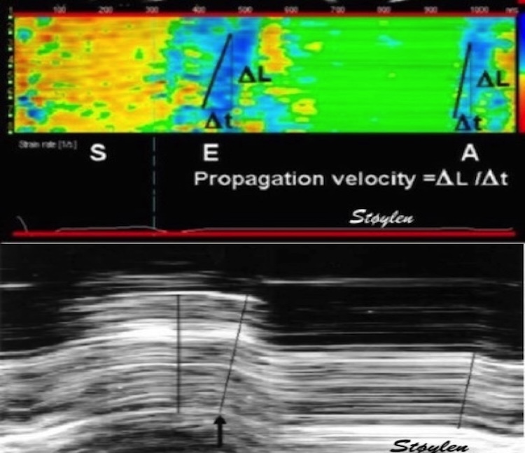
/2 Strain analysis of the LV myocardiumtreats the LV myocardium as ONE object deforming in three dimensions, longitudinal, transmural and circumferential.
The three normal LV strains; longitudinal, circumferential and transmural do not reflect directional fibre shortening, they are simply the three spatial coordinates of the total deformation of the three-dimensional object; the LV myocardium.
the xyz and longitudinal/transmural/circumferential coordinate systems are equivalent. Nobody would talk about three independent functions along the x/y/z axes, so why would it be along the l/t/c axes?
/3 The three strains are interrelated, indicating very little myocardial systolic compression. This means that the main information is carried by longitudinal strain. When an incompressible object deforms along ONE dimension, it has to deform along the other two as well.
When the LV shortens, it has to thicken. Wall thickening pushes both endocardial and midwall circumferences inwards so they shorten. Circumferential strain is the same as diameter shortening.
/4 There is a gradient of circumferential strain from outer to inner circumference. Outer circumferential shortening is the true circumferential fibre shortening, while the deeper circumferential strains are increasingly a function of wall thickening.
/5 All three strains are body size, gender and age dependent. Gender dependency is only due to body size. Both age and body size dependency is highest for longitudinal strain.
All there strains decline with age. Thus, there is no increase in short-axis (circumferential) function to compensate for the reduced longitudinal function, despite preserved EF by age.
• • •
Missing some Tweet in this thread? You can try to
force a refresh
Old misconceptions become as new. A 🧵 A recent paper focusses on pre ejection velocities as a contractility measure. In addition, the authors maintain that these velocities are isovolumic contraction, which they also maintain, is load independent. pubmed.ncbi.nlm.nih.gov/37816446/
All three concepts are wrong. True, the peak contraction velocity (peak rate of force development) occurs before AVO, and thus is afterload independent. But it's not preload independent and thus not a true contractility measure. pubmed.ncbi.nlm.nih.gov/13915199/
🧵 on atrial systole. 1/ Already in 2001, did we show that both the early and late filling phase was sequential deformation propagating from the base to the apex. pubmed.ncbi.nlm.nih.gov/11287889/
2/ This means, both phases consist of a wall elongation wave, generating an AV-plane motion away from the apex. So what are the differences?
3/ Only e’ correlates with MAPSE, so the elastic recoil is finished in early systole, while a’ do not, so atrial systole is a new event, caused by the next atrial contraction. pubmed.ncbi.nlm.nih.gov/37395325/
🧵1/ Sorry, I accidentally deleted the first tweet in this thread, here is a new and slightly improved version. Looking at the physiology of AVC propagation velocity, there are confounders galore, so taking it as a marker of fibrosis, is premature, to put it mildly.
2/ Firstly, The AVC is an event of onset of IVR, i.e at a part of heart cycle with relatively high cavitary and myocardial pressure. This may contribute to wall stiffness, which again may affect (probably increase) wave propagation velocity.
3/ Secondly, This may affect AS patients; who may have a higher wall/cavity pressure at end systole than controls, and thus higher pressure related stiffness.
🧵1/ Looking at the physiology of AVC propagation velocity, there are confounders galore, so taking it as a marker of fibrosis, is premature, to put it mildly.
2/ Firstly, The AVC is an event of onset of IVR, i.e at a part of heart cycle with relatively high cavitary and myocardial pressure. This may contribute to wall stiffness, which again may affect wave prpagation velocity.
3/ Secondly, AS patients may have a higher wall/cavity pressure at end systole than controls, and thus higher pressure related stiffness.
🧵 On early diastole. 1/ It is important to differentiate relaxation and myocyte elongation. Relaxation means tension devolution, due to the removal of Ca, and dissolution of actin/myosin cross bridges. Elongation means volume expansion. They are not simultaneous.
2/ Myoccyte relaxation actually starts during ejection at the time of peak pressure, the decreasing pressure during ejection shows decreasing myocyte tension. pubmed.ncbi.nlm.nih.gov/6227428/
3/ Simultaneously, ejection continues, chiefly due to inertia, until overcome by the Ao-LV pressure gradient, when AV closes. Thus, there is simultaneous myocyte relaxation (tension↓) and volume ↓ (= myocyte shortening). Here is blood flow / myocardial deformation interaction
🧵1/ The E/A fusion in mitral flow with higher HR is well known, normally occurring around HR 100.
2/ also, it should be well known that this occurs because the diastole shortens more with high HR than systole. But why?
3/ In an early study of intervals during exercise, we showed that the RR-interval and DFP, but not LVET shortened in parallel < HR 100. > HR 100 (< RR 600) Both LVET, DFP and RR interval shortened in paralell, but at a slower rate. pubmed.ncbi.nlm.nih.gov/14611824/