Priya Rao, MD Profile picture
May 13, 2020 β€’ 3 tweets β€’ 2 min read β€’ Read on X
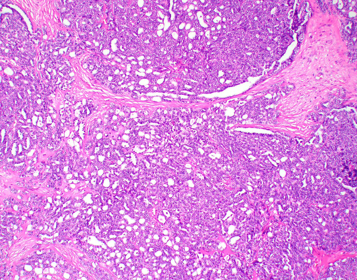
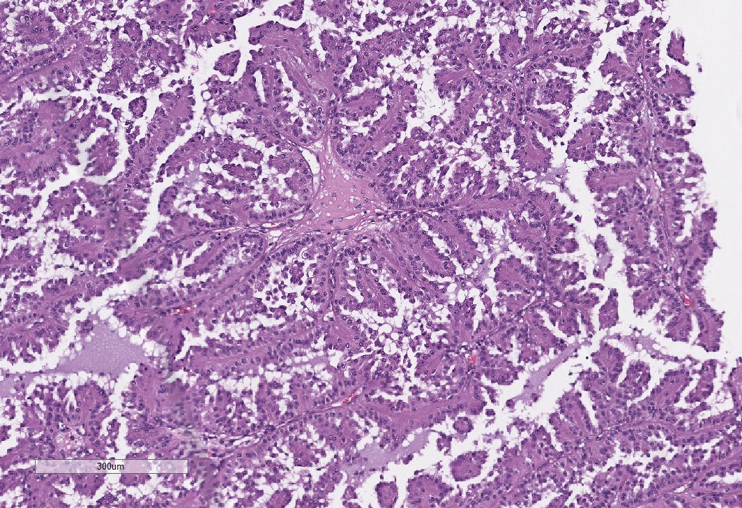
Rib mass in a young male with a renal mass (not biopsied). Morphology was entirely tubulocystic. Thread continued below. πŸ‘‡ #GUpath ImageImage
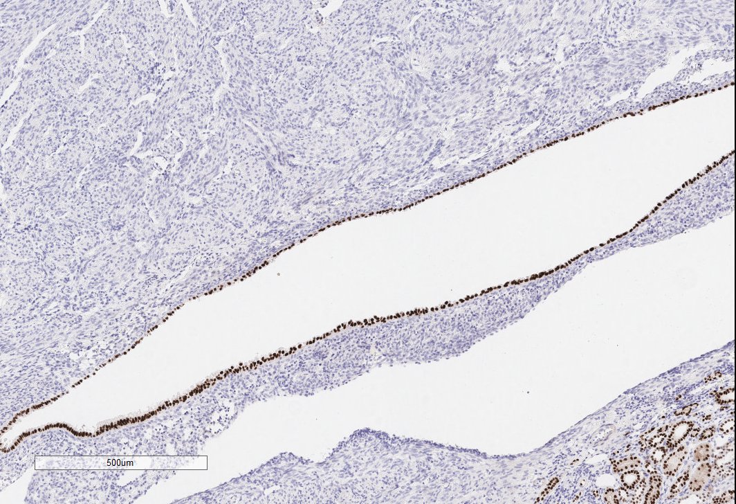
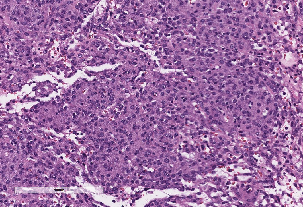
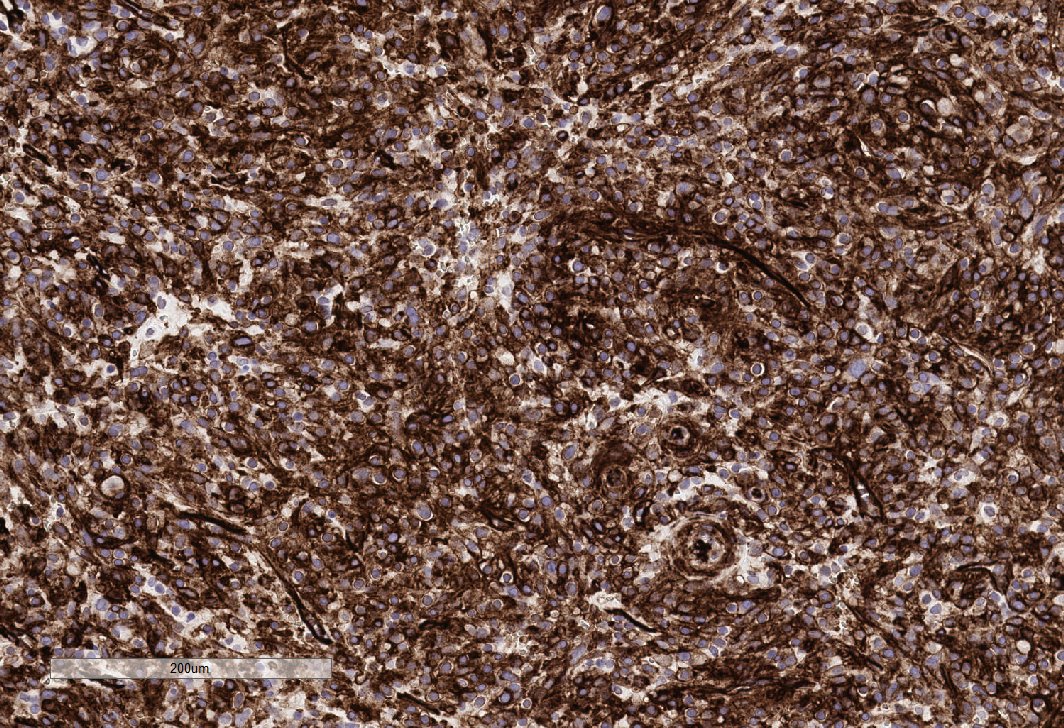
This is what the follow up resection looked like. I received this a few years ago, before we had the FH stain in house. Given the extensive tubulocystic morphology and high grade nuclei, I asked the clinician to test the pt for an FH gene mutation (he was + for the mutation). πŸ‘‡ ImageImageImage
Final diagnosis: HLRCC.

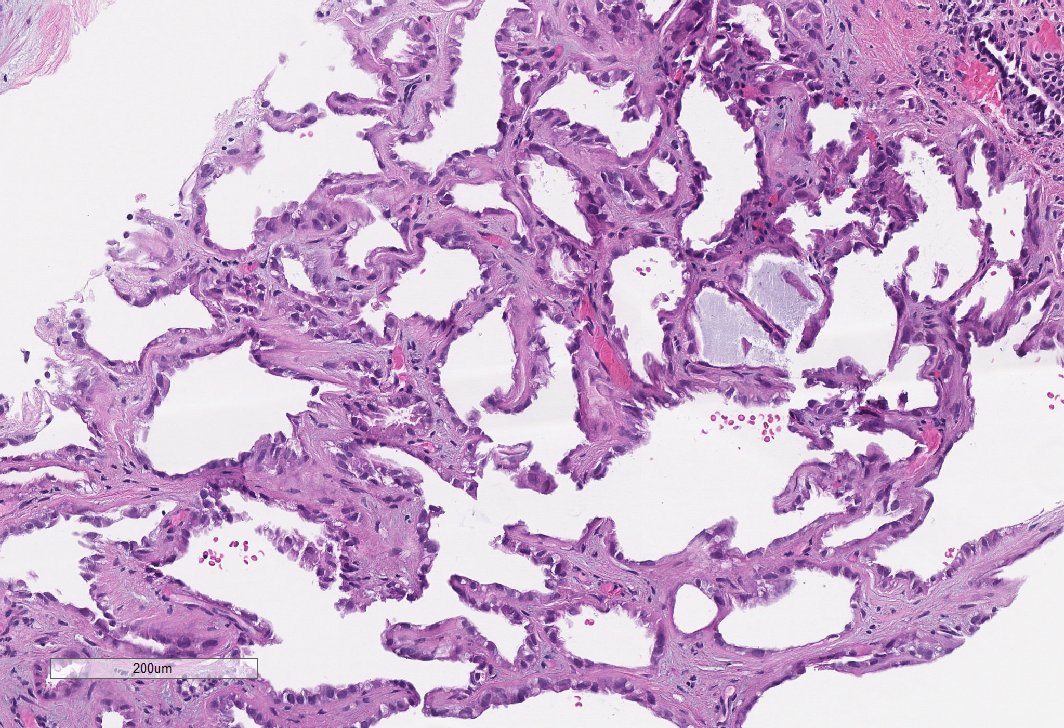
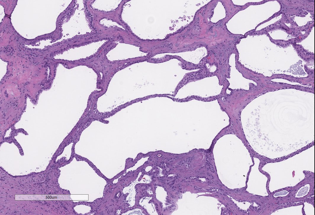
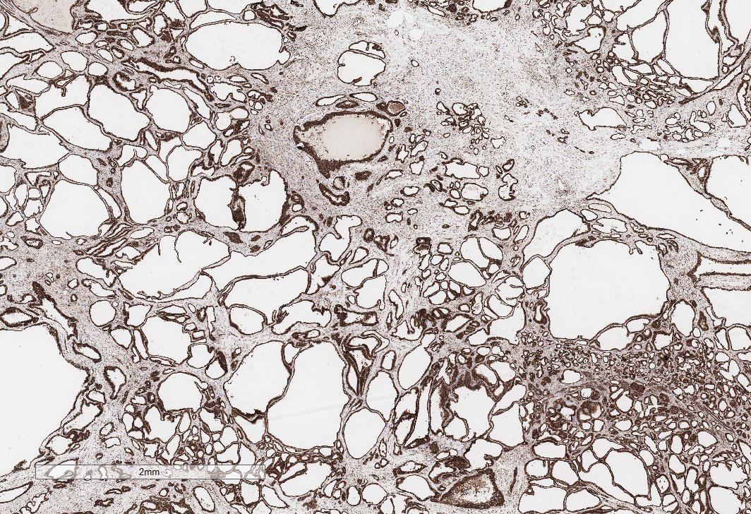
Tumors may be extensively tubulocystic (TC), especially on bx. These are far more common (in my practice) than true TC RCC's. I run the FH stain on all RCCs with TC features.

Bx was signed out as RCC with TC features, clinically exclude HLRCC. #GUpath

β€’ β€’ β€’

Missing some Tweet in this thread? You can try to force a refresh
γ€€

Keep Current with Priya Rao, MD

Priya Rao, MD Profile picture

Stay in touch and get notified when new unrolls are available from this author!

Read all threads

This Thread may be Removed Anytime!

PDF

Twitter may remove this content at anytime! Save it as PDF for later use!

Try unrolling a thread yourself!

how to unroll video
  1. Follow @ThreadReaderApp to mention us!

  2. From a Twitter thread mention us with a keyword "unroll"
@threadreaderapp unroll

Practice here first or read more on our help page!

More from @priyaraomd

Sep 3, 2020
#GUPath twitter brain trust. Posting for a colleague.

37yo man with a renal mass.

Pos for PAX8, keratin. Neg for NE markers, GATA, WT1 and everything else.

We are stumped. Thoughts? Anyone seen something like this before? We thought of adult Wilms’s but IHC doesn’t support. ImageImageImageImage
Tumor is primary to the kidney. He’s older and PAX8 +++ so we didn’t think this was teratoma. I did consider it briefly.
Read 5 tweets
May 15, 2020
Renal bx from a woman in her late 50s, with a renal mass and metastasis. This biopsy is from the kidney. This was a highly infiltrative, high grade tumor with a desmoplastic background. Continued πŸ‘‡ #GUpath ImageImage
The referring institute reported + PAX8, CK7, - GATA3, CK20, TTF1. O/S diagnosis was collecting duct carcinoma.

Upon review by us, we realized that the pt was AA and ordered an INI-1 which was negative.

Diagnosis: Renal medullary carcinoma (confirmed by Hb electophoresis) πŸ‘‡
True CDC of the kidney is extremely rare and is a diagnosis of exclusion. All tumors that look like a high grade adenoca, automatically get an INI-1. (I always exclude Uca and mets.)

RMC is usually a disease in younger pts, but can present at any age. #GUpath @PavlosMsaouel πŸ‘‡
Read 4 tweets
May 14, 2020
Continuing yesterdays theme. Renal bx from a mass in a 55 yo. This one showed more stroma and the nuclei lining the cysts were less "ugly". Continued πŸ‘‡#GUpath ImageImageImage
Mass was resected and was <4 cm and well circumscribed. πŸ‘‡ ImageImageImage
Always run an FH stain (pic) on anything that looks tubulocystic . This was also positive for CK7, P504S and neg for CAIX and CD117.

Final diagnosis: Tubulocystic RCC

Continued πŸ‘‡ Image
Read 4 tweets
May 12, 2020
Incidental renal mass. Stains and diagnosis πŸ‘‡#GUpath ImageImageImage
PAX8 (highlighting epithelium) and HMB45 (stains stromal component). ImageImage
Diagnosis: Angiomyolipoma with epithelial cysts (AMLEC) ER highlights condensed subepithelial stroma below the cyst epithelium. Cystic change may be very focal or extensive. Pay attention, especially on biopsies, for a cyst lining. #GUpath Image
Read 4 tweets
May 3, 2020
Renal tumor in a 19 year old. Stains and diagnosis πŸ‘‡ #GUpath ImageImageImage
CD117 and Synaptophysin (pictures) +. PAX8 - Image
CD34+ (pic) keratin - Image
Read 4 tweets
Apr 30, 2020
I had a colleague from private practice ask me if I ever make a diagnosis of RCC with leiomyomatous stroma (RCCLMS). I thought this would be a good time to share an example. Continued πŸ‘‡ ImageImage
An excellent summary of the molecular findings was published this mth in AJSP by @rajalbshah & @Kiril_T_Can suggests that these are unique tumors that harbor mutations in the TSC, MTOR and ELOC (TCEB) genes and are distinct CCRCC. Diffuse expression of CK7 is key.
These are sporadic counterparts of similar tumors seen in the setting of TSC. I have seen several cases like this and there was not a consensus on what these should be classified as. I like this terminology and will be using this in my practice. cc: @SurenaMatinMD
Read 5 tweets

Did Thread Reader help you today?

Support us! We are indie developers!


This site is made by just two indie developers on a laptop doing marketing, support and development! Read more about the story.

Become a Premium Member ($3/month or $30/year) and get exclusive features!

Become Premium

Don't want to be a Premium member but still want to support us?

Make a small donation by buying us coffee ($5) or help with server cost ($10)

Donate via Paypal

Or Donate anonymously using crypto!

Ethereum

0xfe58350B80634f60Fa6Dc149a72b4DFbc17D341E copy

Bitcoin

3ATGMxNzCUFzxpMCHL5sWSt4DVtS8UqXpi copy

Thank you for your support!

Follow Us!

:(