Great to have our work on bacterial attachment in stent-on-chips published in @micromach_mdpi at mdpi.com/2072-666X/11/4…!
👇summary thread below👇
With @GarethLuTheryn @aliizzzzz94 @AMosayyebi @Center4Biofilm @MiNaTherGroup @uos_bioengsci @ukbiofilms @UoSEngineering 1/7
👇summary thread below👇
With @GarethLuTheryn @aliizzzzz94 @AMosayyebi @Center4Biofilm @MiNaTherGroup @uos_bioengsci @ukbiofilms @UoSEngineering 1/7
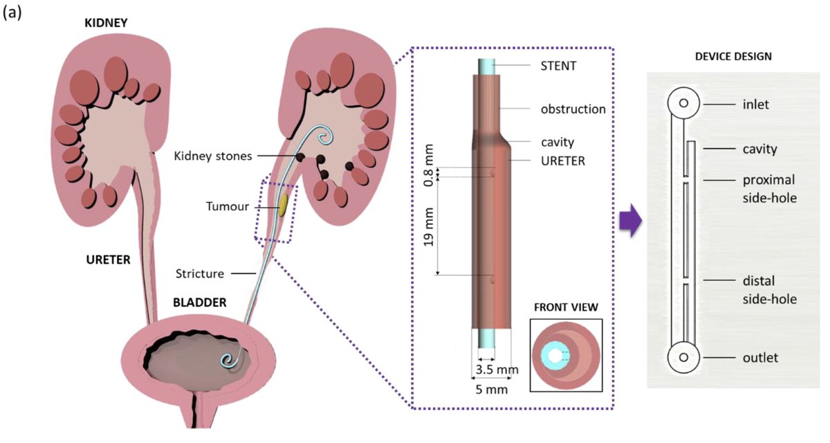
Obstructions of the ureter can be caused by a number of factors and affect the physiological flow of urine from kidney to bladder. To overcome this issue, ureteral stents which have side-holes bypassing urine from ureter to stent and viceversa, are clinically implanted. 2/7 

However, stents are prone to complications leading to infection. We employed microfluidics to mimic flow dynamics in ureteral stents in different obstruction (obs) conditions: 1) unobstructed 2) obs at proximal side-hole 3) obs in between side-holes 4) obs next to side-hole. 3/7 

Obstructions form cavities which are areas of low wall shear stress as shown by CFD simulations. We’ve proven that these are regions where bacteria start depositing (dyed by crystal violet). On the contrary, where there are no cavities, we observe no bacterial attachment. 4/7 

The bacterial attachment, calculated in percentage for the total area of the different designs, was quantified by image analysis of crystal violet dye. We found significant difference between the unobs design compared to the designs with single cavity and double cavity. 5/7 

High-resolution fluorescence images were taken at the site of bacterial attachment using cyto-9 stain. This showed that the number of bacteria is high in the ureter lumen at the cavity region, and decreases when moving to the side-hole and the stent lumen. 6/7 

Based on these results, we proposed an innovative design to produce stents with more side-holes which can help increasing the wall shear stress along the whole system avoiding cavity formation and, therefore, bacterial attachment and infection. 7/7 

#Microfluidics #3Dprinting #FlowDynamics #Imaging #ImageAnalysis #DataAnalysis #Stents #Bacteria #Biofilm #Infection #CFD #Simulations #Ansys #Paper #Published #Bioengineering #Biomedical
@threadreaderapp unroll please
• • •
Missing some Tweet in this thread? You can try to
force a refresh


