A #TWEETORIAL for #RadRes and #MedStudentTwitter
Inspired by the recent @radiology_rsna article: pubs.rsna.org/doi/10.1148/ra…
Important work from our very own @BWHRadEdu @walterfwiggins @AaronSodickson

First some background:
Non-contrast Head CT is the first-line imaging study in trauma.
While most acute hemorrhage can be diagnosed confidently on a CT, small foci of hemorrhage and calcification can be hard to differentiate. This is where dual-energy CT (DECT) can help!
To understand DECT, we have to understand some basic CT physics concepts.
POLL: In diagnostic imaging, photon absorption for high atomic number elements is dominated by what process?
A) Photoelectric effect
B) Compton scattering
C) Rayleigh scattering
D) Pair production
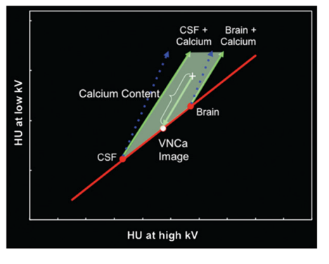
In DECT, x-ray absorption data are gathered from low- and high-energy x-ray spectra to capitalize on the differences in energy-dependent x-ray absorption of different materials.
For example, the mean x-ray energy for a typical 120-kV spectrum is 70 keV.
Since the K edge of Calcium is 4 keV, Ca will result in GREATER increases in attenuation at LOWER kV settings, giving rise to a characteristic slope. A three-material decomposition can be done to separate the component of Ca in every voxel.
Fig from:
pubs.rsna.org/doi/10.1148/rg…

Getting back to the study… the PURPOSE: Quantify the diagnostic performance of DECT versus simulated single-energy CT in the differentiation of small foci of ICH from calcium.
MATERIAL & METHOD
467 consecutive dual-energy unenhanced CTs of the head performed at the @BWHRadiology ED were included if:
1) Hyperattenuating foci > 100 HU
2) Minimum focus diameter > 2.5 mm
3) Binary classification of Ca vs. Hemorrhage was possible.
MATERIAL & METHOD CONTD
All of the single-energy CT scans were read by a #radres and each was classified as:
1) CONFIDENT calcification
2) CONFIDENT hemorrhag
3) INDETERMINATE
REFERENCE TRUTH characterization was made by comparing with a follow-up CT or MRI.
It was classified as HEMORRHAGE:
a) If it was new from 1 month prior
AND IF
b) Perilesional edema on a follow-up image or MRI demonstrated phase signal consistent with blood on SWI
It was classified as CALCIUM:
a) No morphologic features to suggest alternative diagnosis
AND
b) Unchanged from a prior at least 1 month before or after the study or if MRI demonstrated phase signal opposite to blood pool on SWI
The initial assessment made by the #radres was therefore determined as CONCORDANT or DISCORDANT based on comparison with the reference truth.
Hyperattenuating foci were divided into:
A development set of foci, all with CONFIDENT assessment and which were CONCORDANT
AND
Test set of foci which were either INDETERMINATE on initial assessment or with DISCORDANT reference truths.
Quantitative analysis was performed by the #radres placing an ROI centered on the highest attenuation component and recording:
1) Single-energy CT attenuation
2) Calculated attenuation of the VNCa (virtual non-Ca) component
3) Calculated attenuation of the Ca component
The test set was used in a blinded reader study with 2 expert ED Radiologists. In each of the phases, a focus was classified based on the Likert scale as follows:
-2Certain Calcium
-1Likely Calcium
0Uncertain
1Likely Hemorrhage
2Certain Hemorrhage
IN SUMMARY
Dual-energy CT showed high diagnostic performance in the differentiation of small foci of intracranial hemorrhage from calcium and improved diagnostic accuracy and confidence in evaluation of suspected hemorrhage.
unroll


