#Cardiotwitter@fpmorcerf Thread. The end ejection is also a complex series of events, with interaction between the aortic and mitral valves that is reflected in the septal and ring motions.
1/ The determining AVC by Tissue Doppler has been subject to confusion. In the septum, just after the ejection, there is a short negative spike. This was assumed to be IVR, among other things based on the proximity to peak negative dP/dt, which, however only is a proxy for AVC.
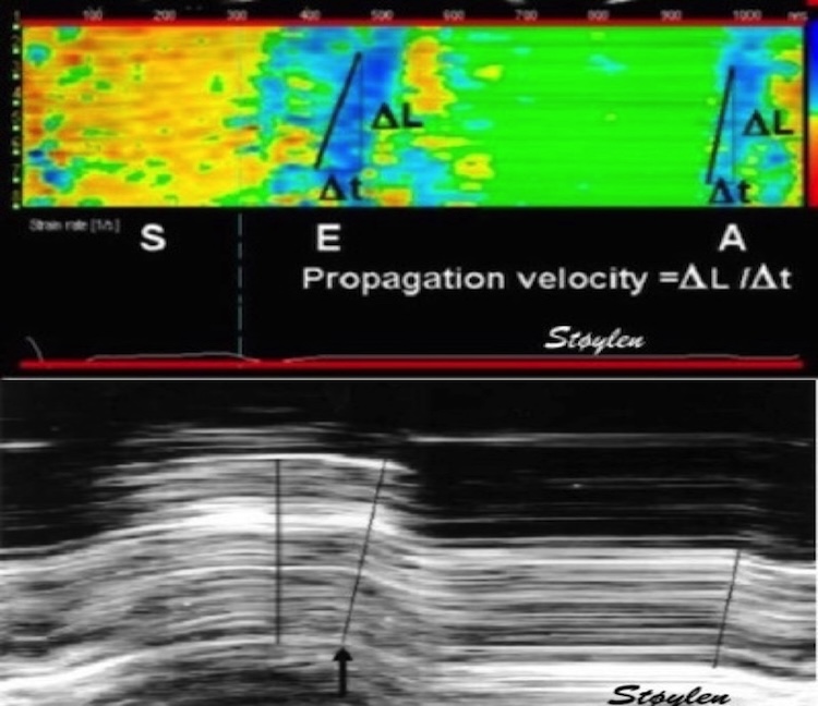
2/ This negative velocity is seen in both septal M-mode, spectral Tissue Doppler, colour TDI, and even as a septal elongation in strain rate. It was visual even in colour TDI of the mitral valve. Thus, AVC was assumed to be at the start of this event.
4/ and it is easily seen by transferring valve motions from Doppler flow to Tissue Doppler recordings, where it seems that this negative velocity is mainly septal.
5/ so just as in the pre ejection spike, this is not IVR, and the true IVR (AVC to MVO) is from the end of the spike to start of mitral flow.
6/ But is this interesting? Yes, firstly for correct measuring of IVRT by using TDI. Secondly, because this in itself gives information about aortic valve closure. Myocyte relaxation, in the meaning of tensjon development, starts at peak LV systolic pressure.
7/ Shortening, continues, under decreasing tension, due to continuing, but decreasing outflow. At end of flow, there is no shortening, but continuing relaxation. This implies elongation with open aortic valve.
8/ The motion of the aortic root in a stationary blood column will in itself be a mechanism for closing the valve, and the valve closure itself be a mechanism for termination of this protodiastolic motion, but also the aortic orifice will "capture) a small volume at end ejection
9/ This has been shown experimentally, pubmed.ncbi.nlm.nih.gov/18606917/, both the volume increase before AVC, and that stenting of the aortic valve abolished the protodiastolic spike.
10/The protodiastolic motion of the mitral ring may be asymmetric, as the open aortic orifice will offer less resistance to the basal motion than the closed mitral valve
🧵 on atrial systole. 1/ Already in 2001, did we show that both the early and late filling phase was sequential deformation propagating from the base to the apex. pubmed.ncbi.nlm.nih.gov/11287889/
2/ This means, both phases consist of a wall elongation wave, generating an AV-plane motion away from the apex. So what are the differences?
3/ Only e’ correlates with MAPSE, so the elastic recoil is finished in early systole, while a’ do not, so atrial systole is a new event, caused by the next atrial contraction. pubmed.ncbi.nlm.nih.gov/37395325/
🧵1/ Sorry, I accidentally deleted the first tweet in this thread, here is a new and slightly improved version. Looking at the physiology of AVC propagation velocity, there are confounders galore, so taking it as a marker of fibrosis, is premature, to put it mildly.
2/ Firstly, The AVC is an event of onset of IVR, i.e at a part of heart cycle with relatively high cavitary and myocardial pressure. This may contribute to wall stiffness, which again may affect (probably increase) wave propagation velocity.
3/ Secondly, This may affect AS patients; who may have a higher wall/cavity pressure at end systole than controls, and thus higher pressure related stiffness.
🧵1/ Looking at the physiology of AVC propagation velocity, there are confounders galore, so taking it as a marker of fibrosis, is premature, to put it mildly.
2/ Firstly, The AVC is an event of onset of IVR, i.e at a part of heart cycle with relatively high cavitary and myocardial pressure. This may contribute to wall stiffness, which again may affect wave prpagation velocity.
3/ Secondly, AS patients may have a higher wall/cavity pressure at end systole than controls, and thus higher pressure related stiffness.
🧵 On early diastole. 1/ It is important to differentiate relaxation and myocyte elongation. Relaxation means tension devolution, due to the removal of Ca, and dissolution of actin/myosin cross bridges. Elongation means volume expansion. They are not simultaneous.
2/ Myoccyte relaxation actually starts during ejection at the time of peak pressure, the decreasing pressure during ejection shows decreasing myocyte tension. pubmed.ncbi.nlm.nih.gov/6227428/
3/ Simultaneously, ejection continues, chiefly due to inertia, until overcome by the Ao-LV pressure gradient, when AV closes. Thus, there is simultaneous myocyte relaxation (tension↓) and volume ↓ (= myocyte shortening). Here is blood flow / myocardial deformation interaction
🧵1/ The E/A fusion in mitral flow with higher HR is well known, normally occurring around HR 100.
2/ also, it should be well known that this occurs because the diastole shortens more with high HR than systole. But why?
3/ In an early study of intervals during exercise, we showed that the RR-interval and DFP, but not LVET shortened in parallel < HR 100. > HR 100 (< RR 600) Both LVET, DFP and RR interval shortened in paralell, but at a slower rate. pubmed.ncbi.nlm.nih.gov/14611824/
🧵 As for MAPSE, we showed in HUNT3 thatpwTDI S' varies between mitral ring sites. LV global S' must be averaged, but we have shown that the difference between mean of septal/lateral and of septal/anterior/lateral/inferior is negligible.
2/ Values are age dependent, and in fact mean of 2 walls was 8.37 cm/s, and of four walls 8.4 cm/s, the difference was statistically significant, but totally un interesting as lower measurement limit of pwTDI is 0.1 cm/s. folk.ntnu.no/stoylen/strain…
3/ But what about diastolic velocities? variation of e' between sites is present as for S', as shown previously in HUNT3. It is common to average lateral/septal, but I haven't found any comparisons between two and four sites, so I looked at that in HUNT3 and found: