WuidQ: Washington University ID Questions Profile picture
Jun 12, 2020 โ€ข 13 tweets โ€ข 6 min read โ€ข Read on X
Part 1 of 2
19M +3 wks fever, confusion. Exam notable๐Ÿ‘‡. Brain MRI +frontal lobe masses. CSF: lymph pleo, low gluc. CSF/blood cx, viral PCRs, Histo/Crypto/Blasto/Cocci(-). IGRA/AFB smear/Cx (-). Brain bx: granuloma, +necrosis, AFB/GMS/Cx(-). No response to Abx. ๐Ÿ‘‰ Part 2 Image
Part 2 of 2
Previously healthy, no PMH exc for recent dx of โ€œskin TBโ€ when he p/w nasal skin lesion ๐Ÿ‘†that showed granuloma w/ (-)AFB/GMS on skin bx. Started on RIPE 2 mos prior to presentation. Lives in rural Minnesota, (-)travel. Given SSx, w/c of the ff is the most likely Dx?
1/11 50% got the right answer! Nice job. A case of Balamuthia mandrillaris.

Balamuthia, one of the free-living ameba pathogenic to humans (like Naegleria & Acanthamoeba). Like Acanthamoeba, causes granulomatous amebic encepahlitis (GAE).

Pic in MCQ from bit.ly/2YNQFjs
2/11 Balamuthia GAE causes brain abscesses, that maybe ring-enhancing & a/w edema/hemorrhage. Patients p/w headache, seizure, confusion, fever. CSF shows lymphocytic pleocytosis, high protein, and low-normal glucose ๐Ÿ‘‰ difficult to distinguish from fungal/TB CNS infection.
3/11 As @RicardoLaHozMD pointed, one very helpful clue to Balamuthia is the presence of a characteristic rash in the central face/nose the usually precedes the onset of GAE by 2 wks to 2 yrs and is almost always visible on exam.
4/11 Note that this characteristic rash is almost always present in cases reported outside of the US (e.g. Peru) bit.ly/2YEDt09. But this is uncommonly reported in the US (only 6% of cases) bit.ly/2CfP4ev .
5/11 Nevertheless, in a patient who p/w brain abscesses, skin exam could give you clues and can lead to early diagnosis (think Nocardia). Rash, usually painless, nodular-plaque in the central face but can also be in the extremities ๐Ÿ‘‡bit.ly/2Y5Quk9 (esp elbow/knees). Image
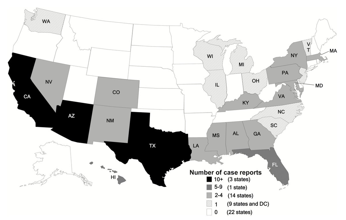
6/11 Balamuthia, reported worldwide. US, common in California, Arizona and Texas๐Ÿ‘‡bit.ly/2CfP4ev. The ecological niche of Balamuthia is not well defined, unlike Naegleria & Acanthamoeba (water). Patients usually report soil > water exposure โ€ผ๏ธ bit.ly/30Ojvmf Image
7/11 Diagnosis is challenging ๐Ÿ‘‰ usually requires brain biopsy. Unlike Naegleria, ameba is not usually seen in CSF. H&E and PAS staining of tissues to see trophozoites & cysts. Requires PCR testing of tissues in most cases (hard to distinguish from Acanthamoeba on microscopy).
8/11 Balamuthia is a fatal disease. Treatment is usually delayed (esp in cases w/ no skin lesion) and ineffective (combination of different antimicrobials including: pentamidine, sulfadiazine, flucytosine, miltefosine).
9/11 Refer to Table ๐Ÿ‘‡ that summarizes the key features of Balamuthia GAE as compared to Acanthamoeba & Naegleria. #highyield @grepmeded Image
10/11 In summary, include Balamuthia in your DDX for brain abscesses of unknown etiology, esp in those w/ concurrent/preceding nasal/central face skin lesion. Closest DDX: fungal and TB. May require brain biopsy, contact health department to help w/ testing.

One more thing...
11/11 I always think that midline face/nose lesions can sometimes help you generalte DDX. See Table ๐Ÿ‘‡ @grepmeded #highyield . A case of Klebsiella rhinoscleromatis was one of the first cases we featured here at @WuidQ 2 years ago ๐Ÿ‘‰ Image

โ€ข โ€ข โ€ข

Missing some Tweet in this thread? You can try to force a refresh
ใ€€

Keep Current with WuidQ: Washington University ID Questions

WuidQ: Washington University ID Questions Profile picture

Stay in touch and get notified when new unrolls are available from this author!

Read all threads

This Thread may be Removed Anytime!

PDF

Twitter may remove this content at anytime! Save it as PDF for later use!

Try unrolling a thread yourself!

how to unroll video
  1. Follow @ThreadReaderApp to mention us!

  2. From a Twitter thread mention us with a keyword "unroll"
@threadreaderapp unroll

Practice here first or read more on our help page!

More from @WuidQ

Jan 6, 2021
38/M w/ progressive loss of scalp, axilla, and chest hairs. Recently dx w/ HIV 6 mos ago when he developed dissem cryptococcosis. He has now been taking TDF/FTC, raltegravir, TMP/SMX, azithromycin, & fluconazole x 6 mos. Drug-induced alopecia is suspected. Most likely culprit?
1/8 Nice job! 52% got the right answer, fluconazole.

In animals/humans, fluconazole has been shown to induce telogen effluvium bit.ly/2MMnF9j, one of the most common causes of nonscarring hair loss (see Table ๐Ÿ‘‡ bit.ly/38rTXyN).

@LParraRod @NNolanMD Image
2/8
Normal hair cycle: anagen (growth) ๐Ÿ‘‰catagen (transformation) ๐Ÿ‘‰telogen (resting) ๐Ÿ‘‰ shedding. Cycle is asynchronous (no mass hair shedding). At any given time, 90% of hair are in anagen, 1% in catagen, 10% in telogen.

@ID_fellows @PBMazi @LeMiguelChavez @gayathri25788
Read 9 tweets
Sep 24, 2020
32/M, h/o HSV encep 1 mo ago (s/p 21 d ACV), on ceftri/metronidazole for sacral OM, p/t ER +delusion, fever, seizure. CSF: WBC 25 (L>N), โฌ†๏ธTP, n/l gluc,(-)HSV. Septic w/u all(-). MRI:
b/l temporal lobe enhancement โฌ†๏ธ from prior. Whch of the ff is the best Tx for this condition?
1/11
The group is split b/n steroids & d/c metronidazole. The answer here is Tx w/ steroids. Indeed, this is a case of autoimmune post-HSV encephalitis (anti-NMDA receptor encephalitis post-HSV). Good job @LemuelNonMD
@LeMiguelChavez @adilrashid83 @Orchid10Tree @KhalafSuha
2/11
Metronidazole-induced encephalopathy is predominated by cerebellar Sx w/ a distinct involvement of the dentato-rubro-olivary pathway on imaging. Weโ€™ve talked about it here before. Refer๐Ÿ‘‡for further discussion
Read 12 tweets
Sep 19, 2020
67/M w/ poor control DM, BPH, +10 d dysuria. T38.1, BP 120/80, +tender R CVA. WBC 14. U/A: 21 WBC, UCx: (-)bacteria, +Candida glabrata (fluc-R) x 2 samples. BCx(-), CT: +prostate hypertrophy. Has had no response to ceftriaxone. Has no Foley cath. Which of the ff is indicated?
1/15
The vote is split b/n micafungin and ampho deoxycholate! Thank you for all your responses!

Although micafungin may be a reasonable option, the correct answer here is ampho deoxycholate.

In this tweetorial, we will talk about Candida UTI and its treatment.
@ID_fellows
2/15
Candiduria can be challenging as it can potentially indicate: colonization, UTI, or candidemia/disseminated infxn.

Candiduria from a clean-voided urine sample is uncommon (<1%); more commonly seen in hospitalized patients w/ an indwelling bladder cath.
Read 16 tweets
Sep 16, 2020
ID Miscellany|physical Exam|Signs|Humanities #idmesh
1/20
๐™๐™€๐™‘๐™€๐™ ๐™‹๐˜ผ๐™๐™๐™€๐™๐™‰๐™Ž: ๐˜ผ ๐™‡๐™Š๐™Ž๐™ ๐˜ผ๐™๐™?

Great! Three quarters find inquiring about fever patterns still useful. We will review some of the most important fever patterns.

@ID_fellows

2/20
For centuries, physicians have relied upon meticulous observations to dx infections. For many years, observation of the fever pattern provided physicians w/ important diagnostic clues. However, the advent of abx & advanced dx & imaging has changed this landscape. #idmesh
3/20
Swift initiation of abx & antipyretics make it impossible to verify historical descriptions of certain fever patterns. Hence, inquiry into fever patterns loses its clinical significance bit.ly/33iXCLs.
Read 21 tweets
Sep 11, 2020
29M w severe persistent asthma p/w recurrent exacerbations despite optimal LABA/intranasal steroids. Abs eos 1250, total Ig E 1500, CT +mucus plugging, central bronchiectasis upper-middle lobes. Originally from Mexico, now in Texas. Which of the ff tests is indicated?
1/10
Great job! The majority got the right answer, allergic bronchopulmonary aspergillosis (ABPA).

Recurrent asthma exacerbations despite optimal asthma therapy & eosinophilia a/w mucus plugging and multilobar central bronchiectasis should raise suspicion for ABPA.
2/10
Aspergillosis, classified as saprophytic (aspergilloma), allergic (ABPA, hypersensitivity pneumonitis, allergic sinusitis), or invasive (pulmonary, other organs).

ABPA: hypersensitivity to A. fumigatus; can also occur from other fungi (referred as ABPM, M for mycosis).
Read 11 tweets
Jul 31, 2020
48M +cirrhosis, underwent routine large volume paracentesis. +Abd fullness, (-)fever, abd pain/tenderness, confusion. Ascitic fluid: light yellow, 100 PMNs, SAAG 1.5, Cx +pan-susc E. coli. WBC 8, Crea 0.8, bili 1.8. Which of the ff is best management for this patient?
1/5
Only 21% got this right: no abx, repeat para in 48H.

The dx of spontaneous bacterial peritonitis (SBP) rests on finding >/= 250 PMNs/mm3 in the ascitic fluid. Most patients with SBP are symptomatic (only 13% with no symptoms bit.ly/3gp5nEU)
2/5
The patient in our case is asymptomatic (no fever, abdominal pain, mental status change ๐Ÿ‘‰most common SBP symptoms) and the ascitic fluid is <250. This is a variant of SBP known as:

โ–ช๏ธMonomicrobial nonneutrocytic bacterascites (MNB)
Read 6 tweets

Did Thread Reader help you today?

Support us! We are indie developers!


This site is made by just two indie developers on a laptop doing marketing, support and development! Read more about the story.

Become a Premium Member ($3/month or $30/year) and get exclusive features!

Become Premium

Don't want to be a Premium member but still want to support us?

Make a small donation by buying us coffee ($5) or help with server cost ($10)

Donate via Paypal

Or Donate anonymously using crypto!

Ethereum

0xfe58350B80634f60Fa6Dc149a72b4DFbc17D341E copy

Bitcoin

3ATGMxNzCUFzxpMCHL5sWSt4DVtS8UqXpi copy

Thank you for your support!

Follow Us!

:(