Asbjørn Støylen 🇳🇴🇩🇰 Profile picture
Sep 20, 2020 18 tweets 5 min read Read on X
1/ Tweetorial. Prompted by a question of "layer strain", I'd like to go into that, as the concept is based on a completely erroneous perception of strain components somehow related to the directional fibre shortening. This is not the case.
2/ The three normal strains are longitudinal, circumferential and transmural (or radial). The relations between all three major strains are explored in the HUNT study: openheart.bmj.com/content/openhr…
3/ Transmural (radial) strain is simply wall thickening, while circumferential strain is fractional circumferential shortening, which, as the circumference is 3.14*diameter, equals fractional diameter shortening.
4/ This means that strains can be measured by calliper measurements, without speckle tracking, although with some assumptions of symmetry.
5/ there is a widespread misconception that circumferential strain represents circumferential fibre shortening. The outer circumferential (and diameter) systolic shortening is 10 - 15%, and represents the true circumferential fibre shortening. pubmed.ncbi.nlm.nih.gov/31673384/
6/ there is a gradient of circumferential shortening with increased numerical values from the outer to the endocardial circumference, the table is from the HUNT study. This has erroneously been described as differential layer function, but is just a function of circular geometry.
7/ As the wall thickens, both midwall and endocardial circumferences moves inwards in response to this, and shortens as a function of this inward motion. This means that circumferential strain is mainly a function of wall thickening.
8/ Midwall circumference is pushed inward by thickening of the outer half of the wall, while endocardial circumference is pushed inward by the whole thickness of the wall, circumferential shortening is numerically highest at the endocardium.
9/ This, is also true of transmural strain. Each layer of the wall towards the endocardium expands into a smaller space (smaller diameter), meaning layers thicken more towards the endocardium. Hence, the midwall circumference moves inwards even relative to the tissue.
But what is wall thickening? As the ventricle shortens, the myocardium, being more or less incompressible, must thicken to conserve volume. Thus, both transmural strain and circumferential strain are strongly related to longitudinal strain.
11/All three strains are the 3D spatial *coordinates* of the total deformation of a single 3d object (LV myocardium), and NOT a function of different fibre directions. All there strain components result from total fibre shortening.
12/ Still, reduced circumferential strain will carry diagnostic and prognostic information, endocardial strain the most, but again, this is simply the result of reduced wall thickening, so it’s the emperor’s new clothes again.
13/ And reduced wall thickening is seen in increased wall thickness.
14/ But what about longitudinal layer strain? It has been described with a gradient, strain being numerically highest in the endocardial layer, and lowest in the outer layer. Is this differential fibre function?
15/ If so, increased shortening in the endocardial slyer, and least in the outer, would give torsion of the mitral ring, which is counterintuitive. To say nothing of the effect on the tricuspid part of the total AV-plane🤯
16/ With only wall thickening, no wall shortening (not compatible with volume conservation, but illustrative), any application tracking wall thickening inwards (e.g. ST), shows shortening, although there is none. And greatest at the endocrdium. Curvature is not a prerequisite.
17/ so differential layer shortening is a function of wall thickening that is tracked by the application, and differential fibre shortening is unlikely as a cause. Again, that means that the midwall and endocardial longitudinal strain is over estimated by tracking.
18/ and again: the three strain components are spatial coordinates of the total 3D deformation, not measures of fibre direction function.

• • •

Missing some Tweet in this thread? You can try to force a refresh
 

Keep Current with Asbjørn Støylen 🇳🇴🇩🇰

Asbjørn Støylen 🇳🇴🇩🇰 Profile picture

Stay in touch and get notified when new unrolls are available from this author!

Read all threads

This Thread may be Removed Anytime!

PDF

Twitter may remove this content at anytime! Save it as PDF for later use!

Try unrolling a thread yourself!

how to unroll video
  1. Follow @ThreadReaderApp to mention us!

  2. From a Twitter thread mention us with a keyword "unroll"
@threadreaderapp unroll

Practice here first or read more on our help page!

More from @strain_rate

Apr 14
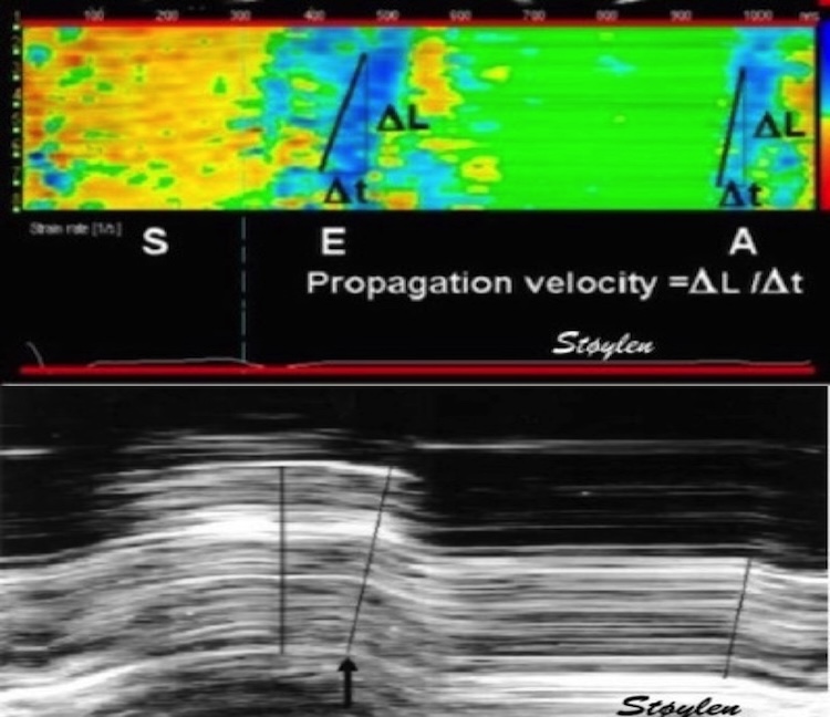
🧵 on atrial systole. 1/ Already in 2001, did we show that both the early and late filling phase was sequential deformation propagating from the base to the apex. pubmed.ncbi.nlm.nih.gov/11287889/
Image
2/ This means, both phases consist of a wall elongation wave, generating an AV-plane motion away from the apex. So what are the differences? Image
3/ Only e’ correlates with MAPSE, so the elastic recoil is finished in early systole, while a’ do not, so atrial systole is a new event, caused by the next atrial contraction. pubmed.ncbi.nlm.nih.gov/37395325/
Read 12 tweets
Apr 10
🧵1/ Sorry, I accidentally deleted the first tweet in this thread, here is a new and slightly improved version. Looking at the physiology of AVC propagation velocity, there are confounders galore, so taking it as a marker of fibrosis, is premature, to put it mildly.
2/ Firstly, The AVC is an event of onset of IVR, i.e at a part of heart cycle with relatively high cavitary and myocardial pressure. This may contribute to wall stiffness, which again may affect (probably increase) wave propagation velocity. Image
3/ Secondly, This may affect AS patients; who may have a higher wall/cavity pressure at end systole than controls, and thus higher pressure related stiffness.
Read 11 tweets
Apr 10
🧵1/ Looking at the physiology of AVC propagation velocity, there are confounders galore, so taking it as a marker of fibrosis, is premature, to put it mildly.
2/ Firstly, The AVC is an event of onset of IVR, i.e at a part of heart cycle with relatively high cavitary and myocardial pressure. This may contribute to wall stiffness, which again may affect wave prpagation velocity.
3/ Secondly, AS patients may have a higher wall/cavity pressure at end systole than controls, and thus higher pressure related stiffness.
Read 7 tweets
Apr 4
🧵 On early diastole. 1/ It is important to differentiate relaxation and myocyte elongation. Relaxation means tension devolution, due to the removal of Ca, and dissolution of actin/myosin cross bridges. Elongation means volume expansion. They are not simultaneous. Image
2/ Myoccyte relaxation actually starts during ejection at the time of peak pressure, the decreasing pressure during ejection shows decreasing myocyte tension. pubmed.ncbi.nlm.nih.gov/6227428/
3/ Simultaneously, ejection continues, chiefly due to inertia, until overcome by the Ao-LV pressure gradient, when AV closes. Thus, there is simultaneous myocyte relaxation (tension↓) and volume ↓ (= myocyte shortening). Here is blood flow / myocardial deformation interaction
Read 17 tweets
Mar 25
🧵1/ The E/A fusion in mitral flow with higher HR is well known, normally occurring around HR 100. Image
2/ also, it should be well known that this occurs because the diastole shortens more with high HR than systole. But why?
3/ In an early study of intervals during exercise, we showed that the RR-interval and DFP, but not LVET shortened in parallel < HR 100. > HR 100 (< RR 600) Both LVET, DFP and RR interval shortened in paralell, but at a slower rate. pubmed.ncbi.nlm.nih.gov/14611824/

Image
Image
Read 8 tweets
Jan 19
🧵 As for MAPSE, we showed in HUNT3 thatpwTDI S' varies between mitral ring sites. LV global S' must be averaged, but we have shown that the difference between mean of septal/lateral and of septal/anterior/lateral/inferior is negligible. Image
2/ Values are age dependent, and in fact mean of 2 walls was 8.37 cm/s, and of four walls 8.4 cm/s, the difference was statistically significant, but totally un interesting as lower measurement limit of pwTDI is 0.1 cm/s. folk.ntnu.no/stoylen/strain…
Image
3/ But what about diastolic velocities? variation of e' between sites is present as for S', as shown previously in HUNT3. It is common to average lateral/septal, but I haven't found any comparisons between two and four sites, so I looked at that in HUNT3 and found: Image
Read 8 tweets

Did Thread Reader help you today?

Support us! We are indie developers!


This site is made by just two indie developers on a laptop doing marketing, support and development! Read more about the story.

Become a Premium Member ($3/month or $30/year) and get exclusive features!

Become Premium

Don't want to be a Premium member but still want to support us?

Make a small donation by buying us coffee ($5) or help with server cost ($10)

Donate via Paypal

Or Donate anonymously using crypto!

Ethereum

0xfe58350B80634f60Fa6Dc149a72b4DFbc17D341E copy

Bitcoin

3ATGMxNzCUFzxpMCHL5sWSt4DVtS8UqXpi copy

Thank you for your support!

Follow Us!

:(