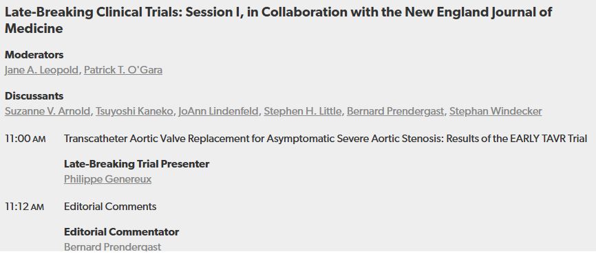
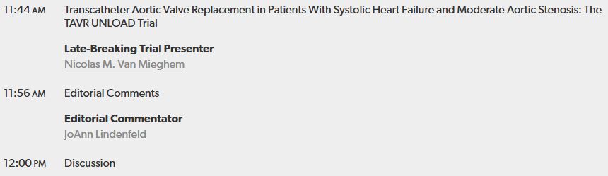
**VALVE CASE OF THE WEEK**
OK, this starts off about valves...but then isn't really about valves...but it's the broader educational point (which is relevant to valves) that I want to make this week...no poll I'm afraid, but as always, comments encouraged! 😁
@BrHeartValveSoc
OK, this starts off about valves...but then isn't really about valves...but it's the broader educational point (which is relevant to valves) that I want to make this week...no poll I'm afraid, but as always, comments encouraged! 😁
@BrHeartValveSoc
In my office doing Admin, lot to get through & a very busy morning ahead. Asked to r/v a TTE for helping determine AS severity. Pt admitted with heart failure, clinically severe AS is all I know at this point. Now, the golden rule in this situation is *review the whole study*...
Not just one or two images.
But I was super-busy, I BROKE MY OWN RULE and just looked at the relevant images. Here's the PW and CW Doppler tracings (Pt in AF)...
But I was super-busy, I BROKE MY OWN RULE and just looked at the relevant images. Here's the PW and CW Doppler tracings (Pt in AF)...


Here's a PSAX view. Images were challenging...
With low velocities and AVA 1.36 I wasn't too worried about severe AS, although did note very low LVOT VTI...but tracing could have been an underestimate due to image quality.
So I said quickly not severe AS, moderate at worst.
With low velocities and AVA 1.36 I wasn't too worried about severe AS, although did note very low LVOT VTI...but tracing could have been an underestimate due to image quality.
So I said quickly not severe AS, moderate at worst.
Next day asked to look again. Colleague convinced clinically AS was severe. So, this time, with more time dedicated to this, I look at the *whole* study, from the beginning, and now something else starts going through my mind! See what you think...here's the PLAX view...
Anything catch your eye? Apart from the monumental LA? Well, I was curious about something so checked out Ap4Ch...👇
What do you think? No BBB on the ECG...
Septum looking a bit funky, right?!
So, next stop...IVC!
What do you think? No BBB on the ECG...
Septum looking a bit funky, right?!
So, next stop...IVC!
IVC measured 25mm with minimal collapse with breathing / sniff...
So, remembering that "a bouncy septum and a dilated IVC is xxx until proven otherwise"....now I decided to check out the other parameters. Some of you know where this is going...
So, remembering that "a bouncy septum and a dilated IVC is xxx until proven otherwise"....now I decided to check out the other parameters. Some of you know where this is going...
MV Doppler shows tall E wave with rapid deceleration time. And check out the TDIs...lateral E' velocity is 13.7! Medial E' is 14.9! They're not normal, they're supranormal! And... they're the wrong way round... that's annulus inversus!!


So, actually, we've got:
-Septal bounce
-Dilated IVC
-Tall MV E wave with short DT
-Supranormal LV E' velocities
-TDI annulus inversus
-Dilated atria (albeit AF)
So now, I'm less worried about AV and I'm wondering...why are there multiple features of constrictive physiology?!
-Septal bounce
-Dilated IVC
-Tall MV E wave with short DT
-Supranormal LV E' velocities
-TDI annulus inversus
-Dilated atria (albeit AF)
So now, I'm less worried about AV and I'm wondering...why are there multiple features of constrictive physiology?!
Turns out a CT scan has commented on unusual thickening of the pericardium!! Cardiac cath awaited.
So, by looking at the *whole* scan, you see the *whole* picture! Sounds so simple, but in the daily hustle of work it's so easy to be tempted into looking at 1 or 2 images only...!
So, by looking at the *whole* scan, you see the *whole* picture! Sounds so simple, but in the daily hustle of work it's so easy to be tempted into looking at 1 or 2 images only...!
Take home message 1: if you're scanning, always think about the numbers you're measuring and images you're seeing...the echo request can't tell you what you may unexpectedly find, so you've got to be alert to expect the unexpected!
Take home message 2: For those asked to review
Take home message 2: For those asked to review
echocardiograms, if it's a "what's the severity of xxx?" question remember to take the time to check all the images...you never know what you may find! 😁
This week's case dedicated to our friend Nicolas @NMerke...hope all is well & look forward to seeing your posts again soon👊
This week's case dedicated to our friend Nicolas @NMerke...hope all is well & look forward to seeing your posts again soon👊
@TheBJCA @BJCA_Women_LTFT @global_wic @WessexSpRs @BSEcho @ASE360 @brwcole @SineadHughes19 @GoughCJ @dorsetcardio @DrChrisMcAloon @coolo_breezo @drstevenwhite @sado_dan @DrWillWatson @jdrwilcox @CardiacJoshi @MayooranShan @drgrahamcole @nieuwoudtrp @TiagoMarta1 @cardiacLucy
@iamritu @rajdoc2005 @HJarrett_MD @rachkataria @RobertoC8a @purviparwani @MadalinaGarbi @JGrapsa @RODRIGOVISCONT1 @RodrigogpLima @shaunrobinson02 @DrDavidWarriner @DrDanAugustine @DrNathan001 @TharushaGunawa4 @echocardiac @echo_stepbystep @nat_echo @The_Echo_Nerd @The_echo_lady
@FGraziani_Grace
@LuciaFGasso
@guyll
@lynne_w12
@LynnGreigMiller @DocStrom
@JohnsonCardio
@JonathanWHinton
@hannahcvimaging
@hannahzr
@BirkhoelzerS
@aayshacader @heartdockumar
@iceman_ex
#accfit #EACVI #cardiotwitter #echofirst
Pls feel free to share / RT!
@LuciaFGasso
@guyll
@lynne_w12
@LynnGreigMiller @DocStrom
@JohnsonCardio
@JonathanWHinton
@hannahcvimaging
@hannahzr
@BirkhoelzerS
@aayshacader @heartdockumar
@iceman_ex
#accfit #EACVI #cardiotwitter #echofirst
Pls feel free to share / RT!
• • •
Missing some Tweet in this thread? You can try to
force a refresh