(3/5) This is because NPM1 mutations cause loss of the nucleolar localization signal and addition of a nuclear export signal, leading to increased protein export from
the nucleus and aberrant accumulation in the cytoplasm
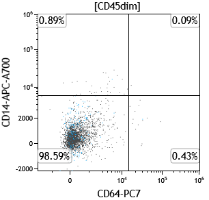
Patient with history of myelomonocytic leukemia post treatment, below is a follow up flow cytometry analysis of his peripheral blood months later, showing an expanded population in the CD45-dim gate (aka "blast-gate") #hemepath #PathTwitter #leusm #MedEd #MedTwitter #MedX @saada_vsaada
This population is negative for CD14, CD33, CD34, C64, CD117, CD123 & HLA-DR expression; However, it shows subset CD13 expression #hemepath #PathTwitter #leusm #MedEd #MedTwitter #MedX
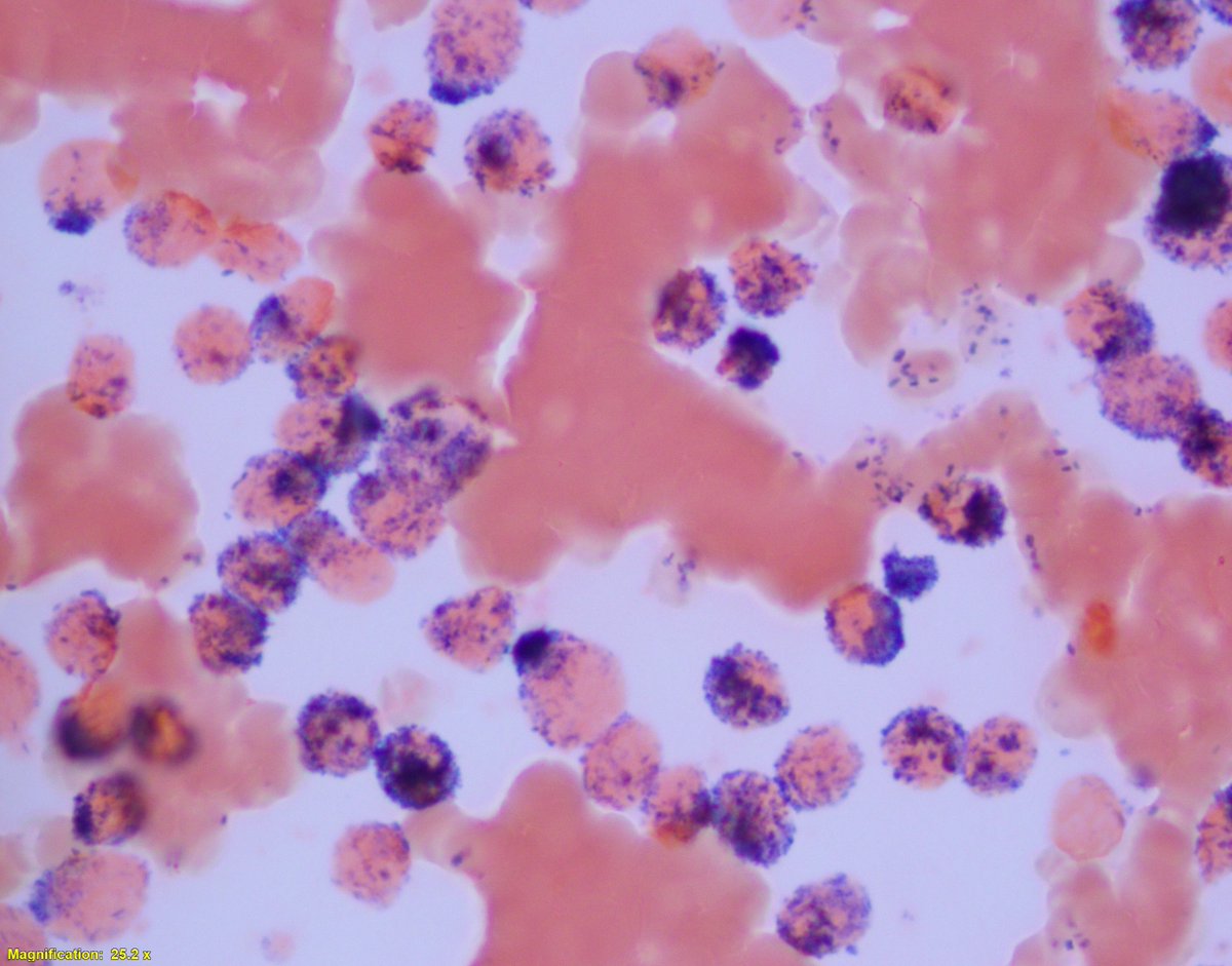
By morphologic examination, no myeloblasts or blast equivalents are identified; However, many cells with the following morphology are appreciated:
Viral infections may show striking T-cell atypia; which in the absence of adequate clinical history, may be worked-up for T-cell lymphoma
Below are pictures from a peripheral blood smear of a patient with an acute viral illness & remarkable lymphocytic atypia #hemepath #lymsm
Flow cytometry analysis confirms the nature of these numerous atypical cells by morphology as lymphocytes, with a predominant CD3-positive T-cell population #hemepath #lymsm #PathTwitter #PathX #SoMe #MedTwitter
This T-cell population consists predominantly CD8+large granular lymphocytes #hemepath #lymsm #PathTwitter #PathX #SoMe #MedTwitter
Patient presented with "pancytopenia, rule out MDS"
Flow cytometry of a limited marrow aspirate revealed a CD20-positive Kappa restricted population that appeared to be negative for CD19 expression #hemepath #lymsm #mmsm #MedTwitter
Morphologic examination showed a hypercellular marrow almost completely involved by a "lymphoid" infiltrate with a low grade morphology
Although by flow cytometry, CD19 was negative, which is odd in a baseline presentation of a low grade lymphoma, CD20 stain was diffusely postive in the infiltrate (below)
An incredibly challenging case of ALK-rearranged poorly differentiated lung adenocarcinoma with diffuse CD30 positivity, mimicking ALK-rearranged Anaplastic Large Cell Lymphoma, first authored by future #hemepath🌟@gibyvgeorge ➡️ #lymsm @Histo_Journalonlinelibrary.wiley.com/doi/10.1111/hi…
Patient presented with a mediastinal mass, composed of sheets of anaplastic cells. Cytokeratin stains along with CD45 & all B-cell & T-cell markers were negative
#hemepath #lymsm #pathtwitter #pathX #molpath #medtwitter #surpath #cytopath #thoracicpath
However, CD30 & ALK1 stains were diffusely & homogeneously positive
High power evaluation: 2 components detetcted, the yellow circle is the CLL/SLL component, and the yellow arrows point at larger pleomorphic cells #lymsm #hemepath #PathTwitter #pathx #surgpath #MedTwitter #bmtsm
In other areas, the background is histiocyte-rich (green arrows) with admixed sacttered large pleomorphic lymphocytes. The first thought one can jump to is: This is classic Hodgkin lymphoma arising from CLL/SLL #lymsm #hemepath #PathTwitter #pathx #surgpath #MedTwitter #bmtsm
CLL/SLL with an inter & peri-follicular architecture ⬇️⬇️
#hemepath #PathTwitter #lymsm #pathX #surgpath
The neoplastic lymphocytes surround germinal centers; If the neoplastic cells had more of a "monocytoid" look, with this perifollicular architecture, this lymphoma could be easily misinterpreted as nodal marginal zone lymphoma, particularly in limited core-nedle biopsy
#hemepath
CD20 stain below highlights the germinal centers in the background (bright expression) #hemepath #PathTwitter #lymsm #pathX #surgpath