⚡️MONTHLY ILLUSTRATIVE CASE⚡️
STATE OF THE HEART BIOPSY
Endomyocardial biopsy is an invaluable yet underutilized diagnostic technique for myocardial disease.
sciencedirect.com/science/articl…
#echofirst #POCUS #FOAMed
@CJC_JCC @Cardio_Girl @SCC_CCS @mustoma
STATE OF THE HEART BIOPSY
Endomyocardial biopsy is an invaluable yet underutilized diagnostic technique for myocardial disease.
sciencedirect.com/science/articl…
#echofirst #POCUS #FOAMed
@CJC_JCC @Cardio_Girl @SCC_CCS @mustoma
Endomyocardial biopsy is typically performed via the right internal jugular using the Seldinger technique and fluoroscopic guidance. This is safely performed with a <1% risk of cardiac complication. 

Fluoroscopic imaging showing endomyocardial biopsy of the RV septum by advancing a bioptome through the RA and tricuspid valve via the right internal jugular vein.
Fluoroscopic imaging showing endomyocardial biopsy of the RV septum by advancing a bioptome through the RA and tricuspid valve via a long sheath in the left internal jugular vein.
Video 3. Fluoroscopic imaging showing endomyocardial biopsy of the RV septum by advancing a bioptome through the RA and tricuspid valve via a long sheath in the right femoral vein.
The primary indications for endomyocardial biopsy are myocarditis, infiltrative cardiomyopathy, and surveillance of heart transplant rejection.
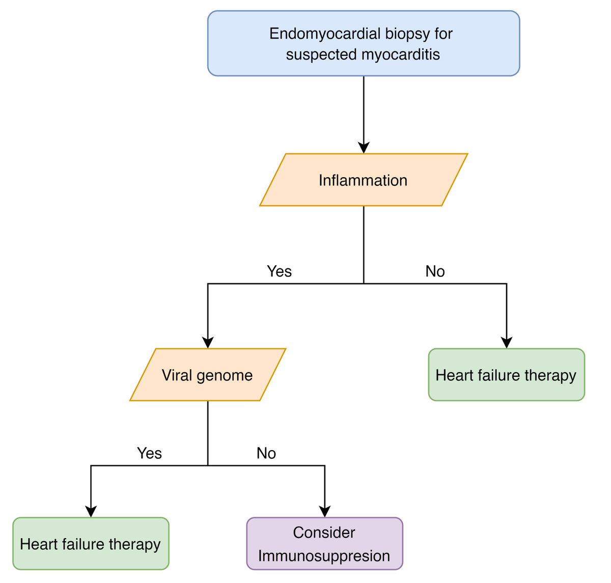
Recommendations for endomyocardial biopsy for unexplained acute cardiomyopathy include high-risk features that may suggest autoimmune myocarditis.
* Including giant-cell myocarditis, acute necrotizing eosinophilic myocarditis, and, if it impacts treatment, ICI myocarditis.
* Including giant-cell myocarditis, acute necrotizing eosinophilic myocarditis, and, if it impacts treatment, ICI myocarditis.

Simplified algorithm for the use of endomyocardial biopsy in the workup of infiltrative cardiomyopathies. 

Cardiac biopsy for tumours may be reasonable if: 1) a non-biopsy diagnosis or non-cardiac-biopsy diagnosis is not possible, and 2) tissue diagnosis will alter management, and 3) an experienced operator is available to perform cardiac biopsy with a high chance of success.
Echocardiographic guidance is used to guide the biopsy of a very large right ventricular tumour.
So please consider endomyocardial biopsy in your diagnostic toolset, particularly for sick patients with myocarditis.
I hope you like this month's illustrative case. Follow and share if you enjoyed. More to come!
OTHER CASES:
LA-to-Aorta LVAD:
Pericardial Waffle:
Aortic Thrombus:
Constriction:
OTHER CASES:
LA-to-Aorta LVAD:
https://twitter.com/OKiamanesh/status/1330716828629303297
Pericardial Waffle:
https://twitter.com/OKiamanesh/status/1302045240422006785
Aortic Thrombus:
https://twitter.com/OKiamanesh/status/1287536558632046592
Constriction:
https://twitter.com/OKiamanesh/status/1267632487406219265
ping!
@leaharper1
@dr_benoy_n_shah
@ArgaizR
@Nmerke
@Iceman_ex
@RJonesSonoEM
@RaynerHartleyMD
@Thind888
@msiuba
@FH_Verbrugge
@ThinkingCC
@fpmorcerf
@AndrewJSauer
@Wormsy10
@YasMoayedi
@leaharper1
@dr_benoy_n_shah
@ArgaizR
@Nmerke
@Iceman_ex
@RJonesSonoEM
@RaynerHartleyMD
@Thind888
@msiuba
@FH_Verbrugge
@ThinkingCC
@fpmorcerf
@AndrewJSauer
@Wormsy10
@YasMoayedi
pong!
@NephroGuy
@NephroP
@oscaar84
@DrBerticMia
@PulmCrit
@dramcarrillo9
@VatsalTrivediMD
@MSharifpourMD
@katiewiskar
@UAlberta_Sono
@drshahrul80
@EmergencyEcho
@AlbertoAOviedo
@AnishRMitra
@JdBapttiste
@NephroGuy
@NephroP
@oscaar84
@DrBerticMia
@PulmCrit
@dramcarrillo9
@VatsalTrivediMD
@MSharifpourMD
@katiewiskar
@UAlberta_Sono
@drshahrul80
@EmergencyEcho
@AlbertoAOviedo
@AnishRMitra
@JdBapttiste
• • •
Missing some Tweet in this thread? You can try to
force a refresh


