It is often said that Marie Skłodowska-Curie died of "aplastic anemia." Try Googling it; you'll find many hits. But I am not so sure. She died on July 4th, 1934, at a sanatorium called Sancellemoz, in Passy, Haute-Savoie, France, after a long illness. #aplasticanemia#MDS /1
The 1937 biography by her younger daughter Ève describes her final illness, including a consultation at Sancellemoz (postcard) by a "Professor Roch." That would have been Maurice Roch, Regent of @UNIGEnews & father of famous Alpinist André Roch who planned Aspen, Colorado./3
Here is how the daughter's biography describes that consultation. Mention is made of fevers and blood tests - rapidly falling WBC & RBC counts - and that X-rays were done. (The last thing she needed: more radiation!). Diagnosis: "Pernicious anaemia in its extreme form." /3
It is unclear if Mme. Sklodowska-Curie underwent a marrow biopsy. The @nytimes ran her obituary the following day, and stated her cause of death as "a form of pernicious anaemia". (Interesting to see @Wimbledon 🎾 scores next to the obituary - it was July 4th week after all.)/4
Aplastic anemia was well recognized by 1934. It had first been described in 1888 at autopsy in a 21 year-old pregnant woman named Hedwig S. with “strikingly hypocellular” marrow - by Paul Ehrlich at Charite in Berlin, in this case report./5
The term "aplastic anemia" is usually said to have been coined in 1904 by this guy: Anatole Chauffard in Paris. The key paper was "Un cas d’anémie pernicieuse aplastique" in the Bulletin Soc Med Hop Paris, 21 (1904): 303f. But... notice the "pernicious" in the paper's title./6
Richard Clarke Cabot in Boston (depicted - he started the clinicopathological case series in @NEJM) summarized 24 cases of aplastic anemia the 1908 version of text, emphasizing hypocellularity. If Mme Curie had #aplasticanemia we might have expected Roch to give that diagnosis./6
(BTW: back then people reported *original* research in textbooks! Osler's 1892 first edition of his Principles and Practice text is packed with his own unpublished observations. Now, of course, textbooks are so long in gestation that anyone who tried that would get 'scooped'.)/7
In contrast to aplastic anemia, myelodysplastic syndromes were not yet well described in 1934. "Refractory anemia" term dates to a 1938 paper by the notorious C.P. "Dusty" Rhoads. I summarized some of that history in a 2012 Leukemia Research paper./8 sciencedirect.com/science/articl…
Since pernicious anemia is associated with blood/marrow cell dysmorphology, as in these 2 @ASH_hematology image bank photomicrographs, and the megaloblastic changes in pernicious anemia overlap with findings in #MDS, many early MDS cases were mistaken for pernicious anemia./9
Aplastic anemia induced by ionizing radiation is often (albeit not always) the result of a single large myeloablative dose. Therapy- or exposure-related MDS, in contrast, has a different epidemiology and may result from lower dose chronic exposure./10 ashpublications.org/hematology/art…
Mme. Curie died at age 66 & had many years of lower-dose exposures to various radioisotopes./11 history.aip.org/exhibits/curie…
Already in 1920 the health effects of radium in particular were well recognized. Mme. Curie wrote to her sister Bronya in 1920, "“Perhaps radium has something to do with these troubles, but it cannot be affirmed with certainty.” "Radium Girl" lawsuits began in the 1920s./12
So while it is not impossible for Marie Skłodowska-Curie to have died of aplastic anemia, as the Nobel Foundation tweeted yesterday, MDS, or another chronic myeloid neoplasm (MF is unlikely given lack of mention of a spleen), or even actue leukemia are more likely. /End
• • •
Missing some Tweet in this thread? You can try to
force a refresh
What is “Bloodburn”? In the @starwars Universe, this mysterious chronic hematologic condition led Greer Sonnel - Senator Leia Organa’s chief of staff - to quit spaceship racing. #HematologyTweetstory 36: hematologic changes from space travel, in fantasy & reality. Image:@NASA/1
First, some sci-fi fun. #StarWars fandom source “Wookipedia” (@WookOfficial, source of below image) tells us Bloodburn is a “rare, chronic, and often terminal illness of the blood that befell (often younger) starship pilots”. Symptoms include fevers... /2 starwars.fandom.com/wiki/Bloodburn
Bloodburn is incurable, but usually manageable with good diet, hydration, rest, & “hadeira serum” injections (the serum itself can be harmful). The pathophysiology of Bloodburn is unclear. The “burn” part suggests radiation mediated-injury, but maybe just refers to the fevers?/3
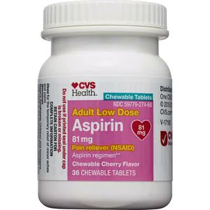
Aspirin continues to be the most widely used anti-platelet agent, 125 years after its synthesis. But where did it come from - and why do we give it in such weird doses (e.g. 81, 162 & 325 mg) – at least in the United States? #HematologyTweetstory 35 will answer these questions./1
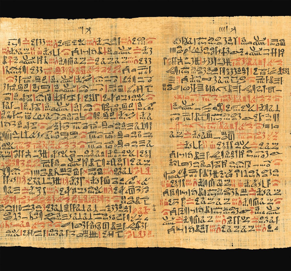
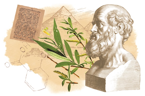
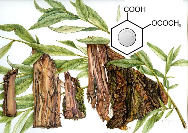
Some lucky ancient person serendipitously discovered that willow bark & leaves relieved pain. Hippocrates used tea made from willow leaf to ease childbirth, while the Egyptian Ebers papyrus (~1500 BCE) mentions willow for aches and pains. (Images: Sermo/Pharmaceutical Journal)/2
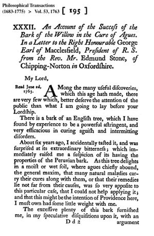
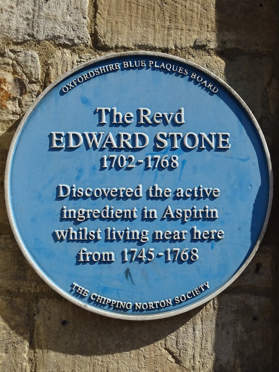
In 1763, @royalsociety published a study of dried willow bark for rheumatism, submitted by Edward Stone (1702-1768), a vicar from Chipping Norton in the Cotswolds & fellow @WadhamOxford. Back then a lot of “natural philosophy” (early science) was done by Anglican clergy./ 3
#HematologyTweetstory 34: Vitamin K. This tale includes 2 larger-than-life characters, self experimentation, & bloody cows. Also, yours truly was once *so* dedicated to hematology history that he drove to rural Wisconsin to search local property records related to this story.😉/1
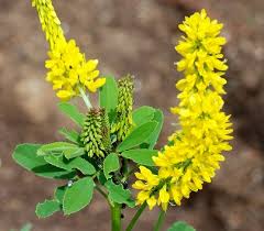
Melilotus: a genus of grassland plants originally from Eurasia, also known as “sweet clover” because of a vanilla-like scent (though the taste is bitter). Sweet clover was first brought to the US & Canada during the Colonial period, and became a useful farm animal feed./2
In the winter of 1921 - a particularly damp season across the upper Midwest - farmers from Wisconsin to the Dakotas, from Ontario to Alberta, had cattle bleed to death. Many calves born the following spring were stillborn & deformed, as if they'd been exposed to a teratogen./3
#HematologyTweetstory 33: hemoglobin variants, often said to be the most common single-gene genetic disorders in humans. “Disorders” is not entirely accurate, as many variants are clinically silent. We’ll focus on hemoglobinopathies; thalassemias are a story for another time./1
I got interested in this ~20 years ago & wrote a paper in 2001 @MayoProceedings about RBC disorders we'd incidentally noted in some of the many patients we saw @MayoClinic from the Middle East (esp. prior to 9/11). I then went to @MRC_WIMM in Oxford to a globin lab for 2 years./2
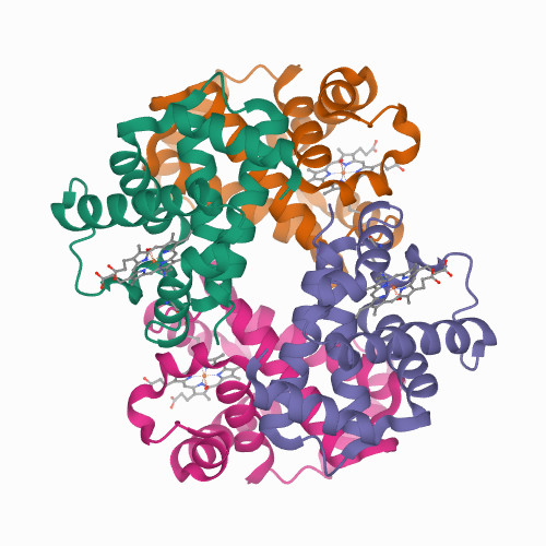
First, a quick run-through of the normal hemoglobins (image source: Hoffbrand and Steensma, Essential Haematology, 8th edition). Already in the 19th century it was recognized that there was more than one type of human hemoglobin. /3
#HematologyTweetstory 32: lymph nodes with names. There’s also a major personal announcement in this thread. We each have lots of lymph nodes: an estimated 500-600 (Image: @MayoClinic). Like stars, they cluster. (Did you ever think of your axilla as a lymphatic “galaxy”?)🙂/1
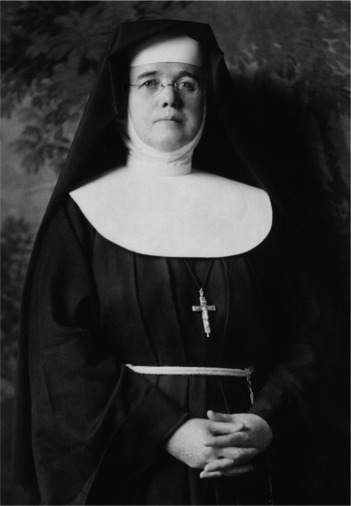
One of the best known eponymous nodes is the “Sister Mary Joseph nodule”, named after the gifted woman born Julia Dempsey (1856-1939), who was Dr Will Mayo’s scrub nurse @MayoClinic and, as a Sister of Saint Francis, directed St Marys Hospital in Rochester, MN for 46 years./2
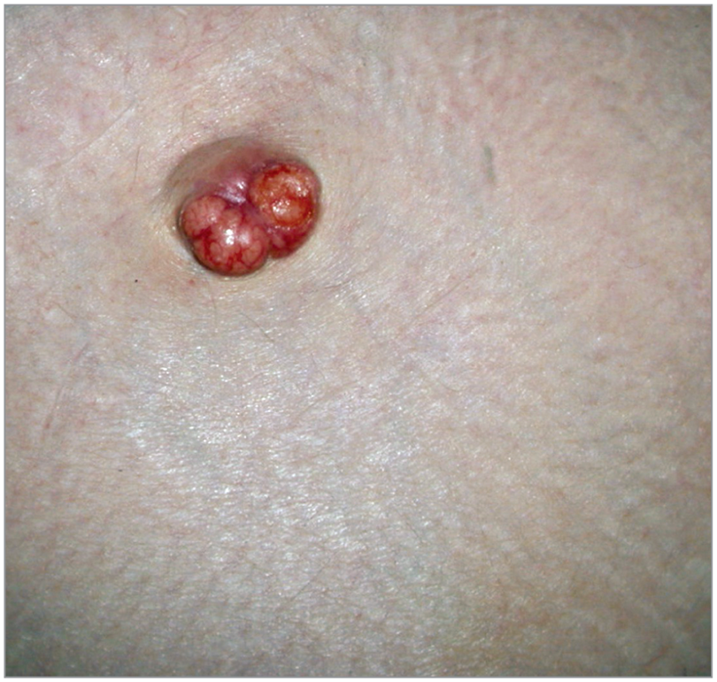
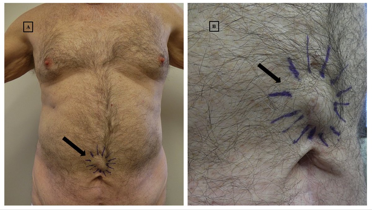
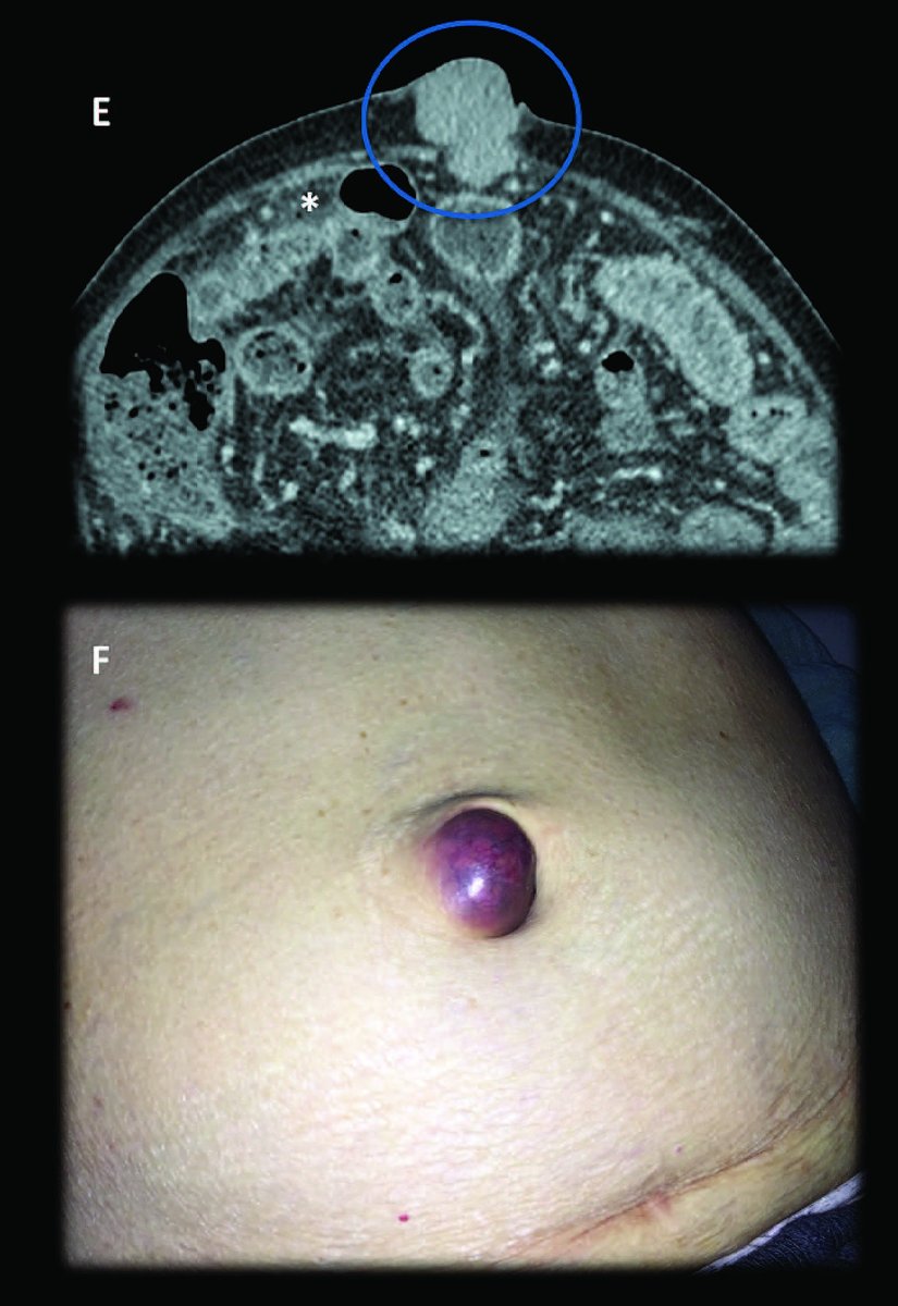
When she scrubbed abdomens before laparotomies, Sister Mary Joseph noticed that whenever there was a firm mass near the umbilicus, the patient turned out to have cancer. Will Mayo published an article in 1928 @MayoProceedings about this – he called it “pants button umbilicus.” /3
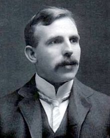
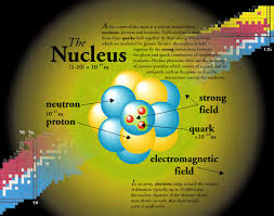
Here’s a thread about the #nucleus… no, Professor Ernest Rutherford, not the atomic nucleus that you discovered with your alpha particles back in 1911.😉 This is about *cell* nuclei and all their weird and wonderful forms, in blood cells and beyond. #HematologyTweetstory 31! /1
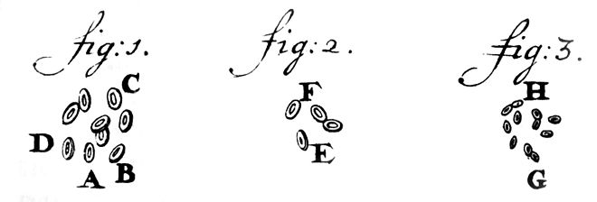
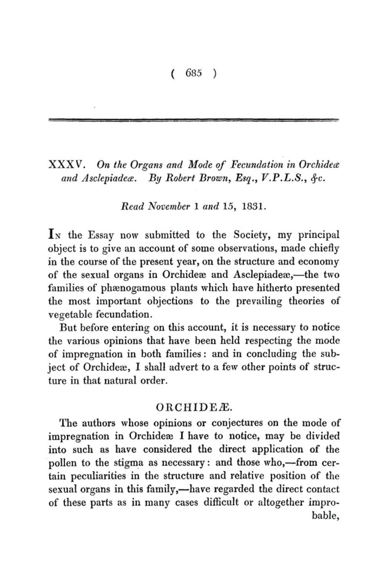
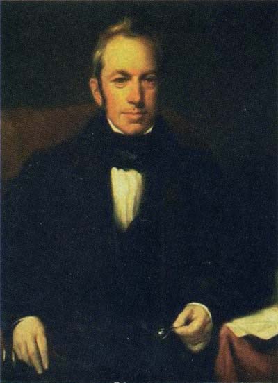
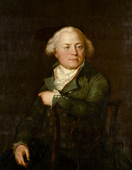
Cell nuclei were first drawn by Dutch microscopy pioneer Antonie van Leeuwenhoek circa 1719 (pictured), and discussed as distinct structures in 1804 by botanist Franz Bauer (below with green jacket), then clearly described in 1831 by botanist Robert Brown (below, with bowtie)./2
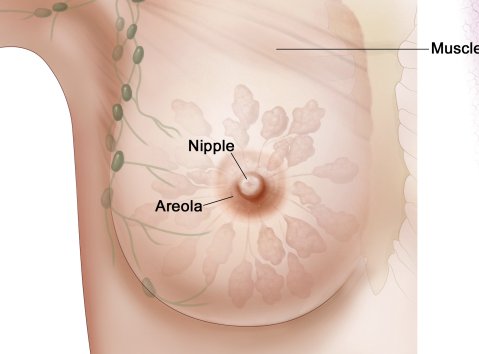
Brown first called the nucleus the “cell areola” – which suggests he may have observed nucleoli as well, although he didn't mention them (they would have been just at the limit of his microscope's resolving ability). Probably a good thing that term didn’t stick, though.../3