Dr John B. Profile picture
Sep 20, 2021 24 tweets 7 min read Read on X
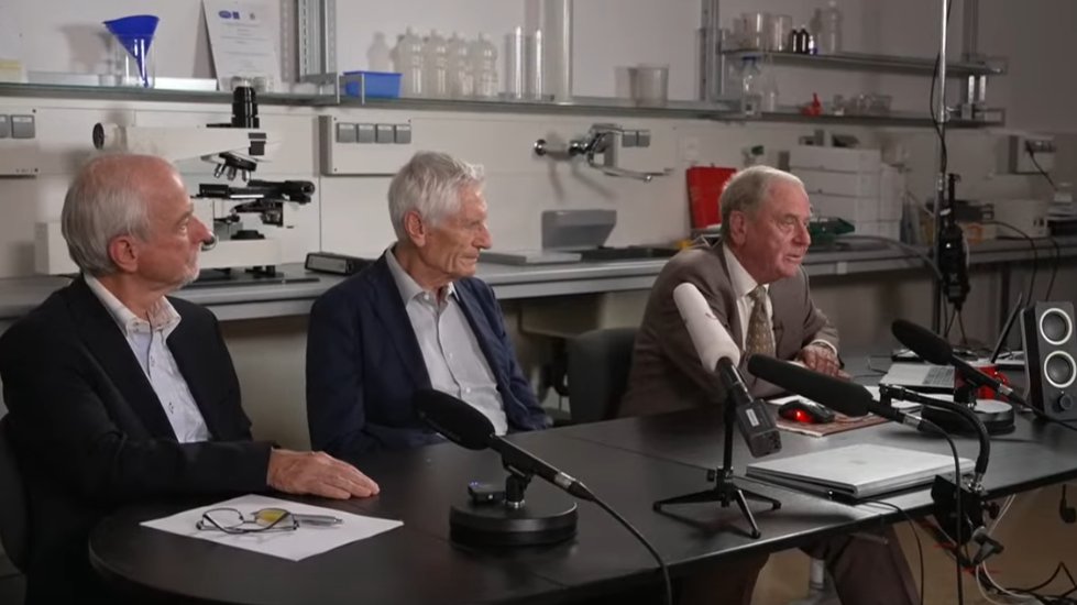
(1/n) Today there was a press conference from the pathological institute in Reutlingen, Germany. The pathologists Prof. Dr. Arne Burkhardt and Prof. Dr. Walter presented the results of the autopsies of eight people who died after COVID19 vaccination.
#CovidVaccine Image
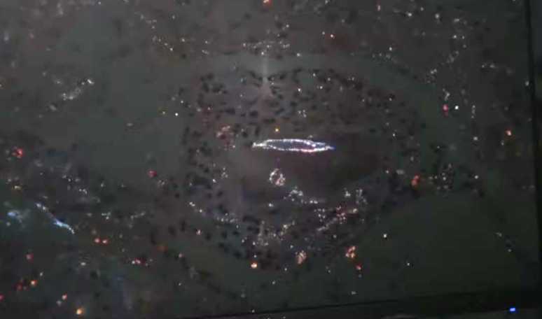
(2/n) In addition, new microscopic analyses of an Austrian research group on the ingredients of the vaccines were presented.
Video: (in German)
Here is a summary of the highlights.
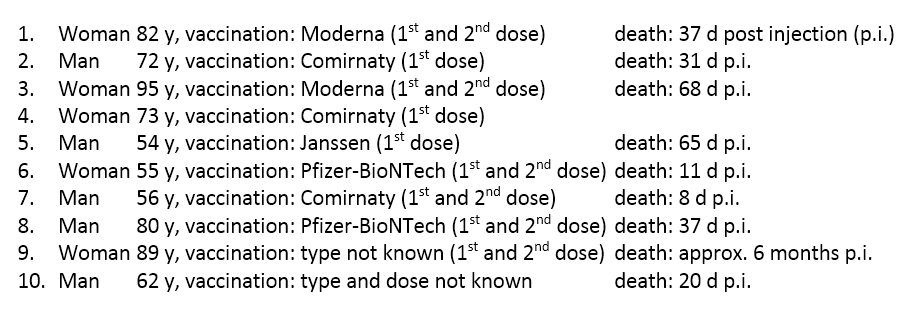
(3/n) 10 autopsies, 8 evaluations.
(Slide text translated into English) Image
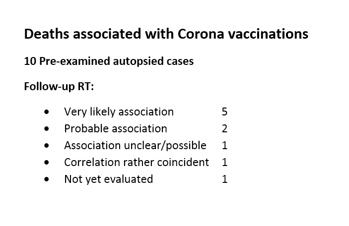
(4/n) Summary.
(Slide text translated into English) Image
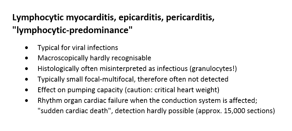
(5/n) Key findings.
(Slide text translated into English) Image
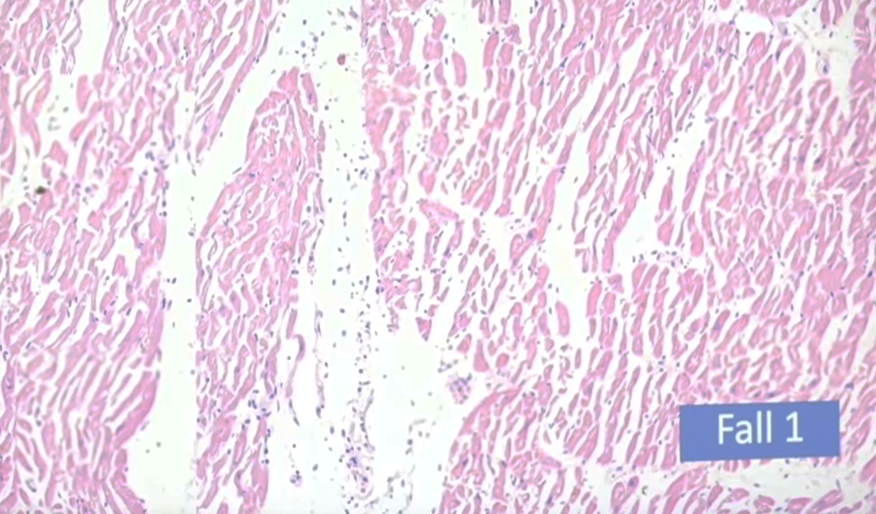
(6/n) Tissue with lymphocytic infiltration.
(Blue dots: lymphocytes). The tissue is inflamed.
Example 1. Image
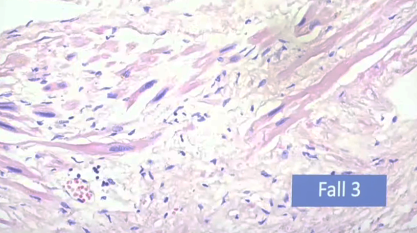
(7/n) Tissue with lymphocytic infiltration.
(Blue dots: lymphocytes). The tissue is inflamed. The muscle fibres are destroyed.
Example 2. Image
(8/n) Tissue with lymphocytic infiltration.
(Blue dots: lymphocytes). The tissue is inflamed.
Example 3. Image
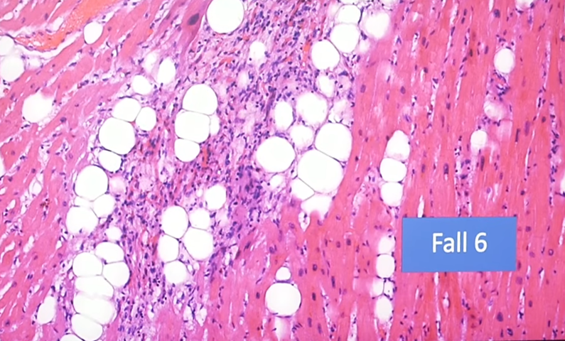
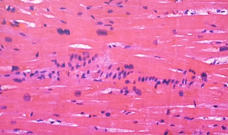
(9/n) Tissue with lymphocytic infiltration.
(Blue dots: lymphocytes). The tissue is inflamed.
Lined up lymphocytes.
Example 4. Image
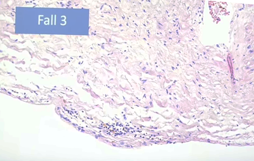
(10/n) Tissue (epicardium) with lymphocytic infiltration.
(Blue dots: lymphocytes). The tissue is inflamed.
Example 5. Image
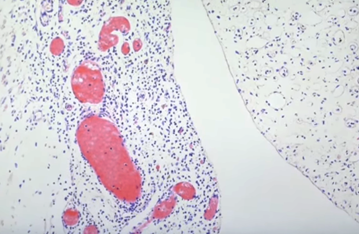
(11/n) Tissue (epicardium) with lymphocytic infiltration.
(Blue dots: lymphocytes). The tissue is inflamed.
Example 6. Image
(12/n) Alveolitis with lymphocytic infiltration.
(Blue dots: lymphocytes). The tissue is inflamed.
Example 7. Image
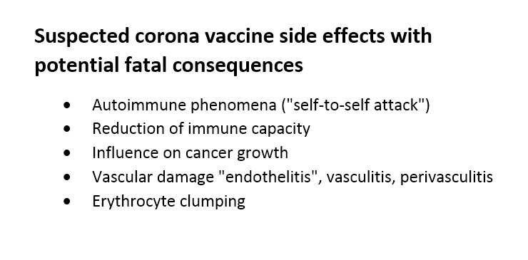
(13/n) Suspected side effects: Image
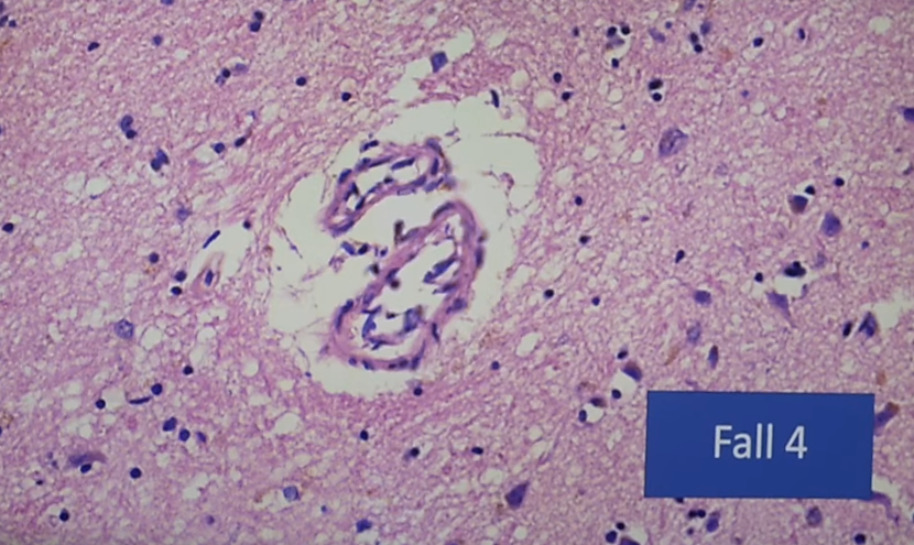
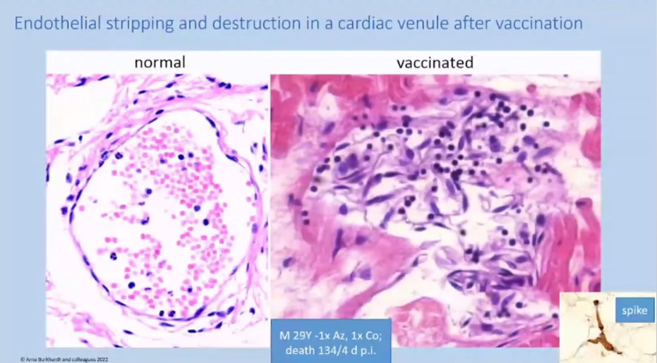
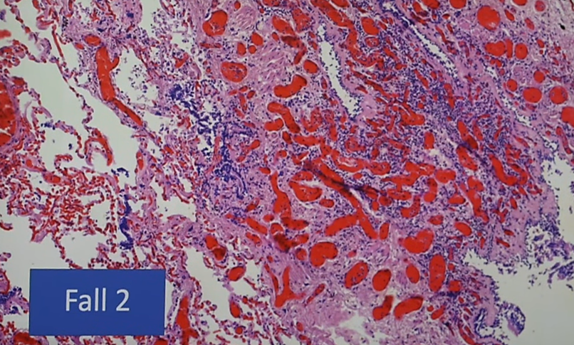
(14/n) Detachment of endothelial cells from the vessel wall.
Example 1. Image
(15/n) Detachment of endothelial cells from the vessel wall.
Example 2. Image
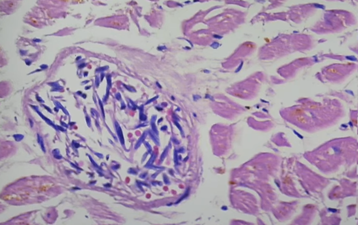
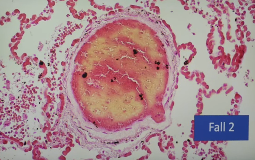
(16/n) Erythrocyte clumping inside a vessel. Thrombosis.
Example 1. Image
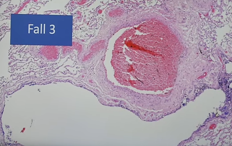
(17/n) Erythrocyte clumping inside a vessel. Thrombosis.
Example 2. Image
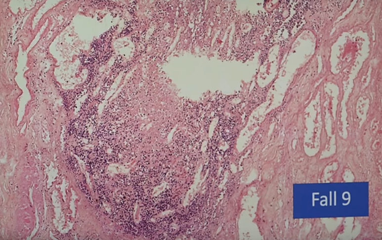
(18/n) Vessel with lymphocytic infiltration.
(Blue dots: lymphocytes).
Example 1. Image
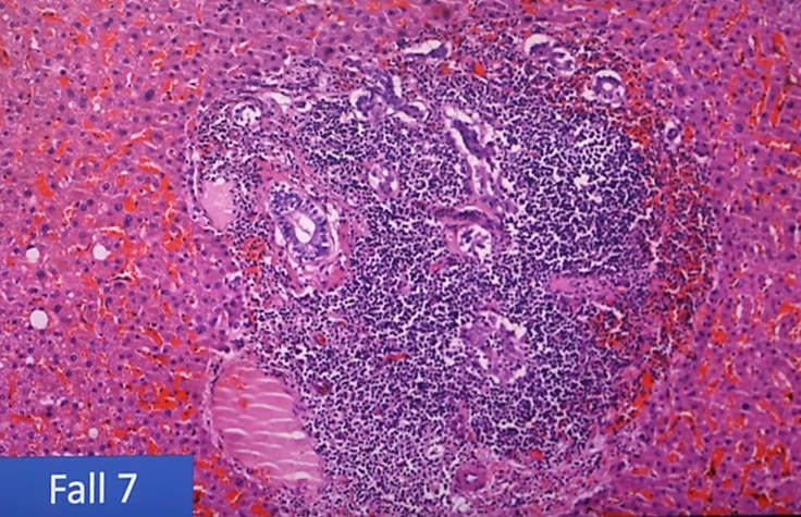
(19/n) Vessel with lymphocytic infiltration.
(Blue dots: lymphocytes).
Example 2. Image
(20/n) Foreign bodies, contaminants, adjuvants in the vaccine.
Microscopic examination of particles of unknown nature.
Example: Image
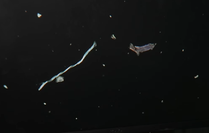
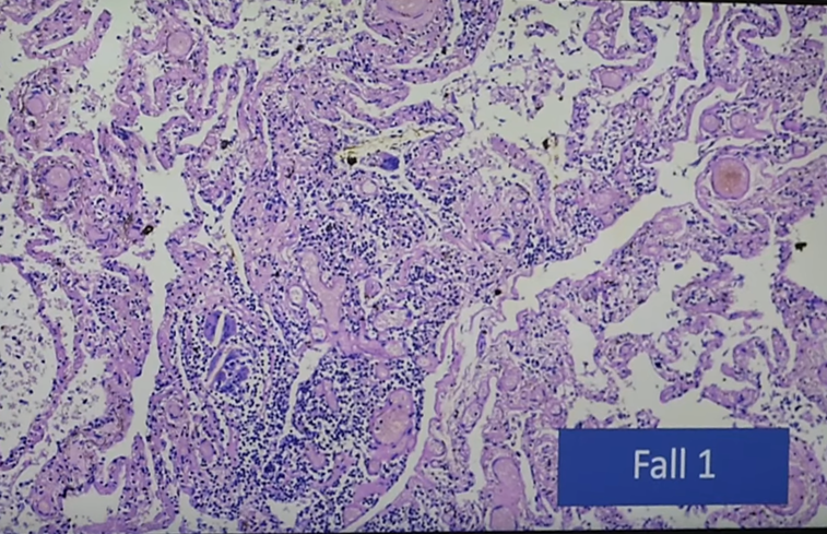
(21/n) A foreign bodies in inflamed lung tissue.
Similar to bone marrow embolism after bone fracture.
Inflammatory giant cells are visible. Image
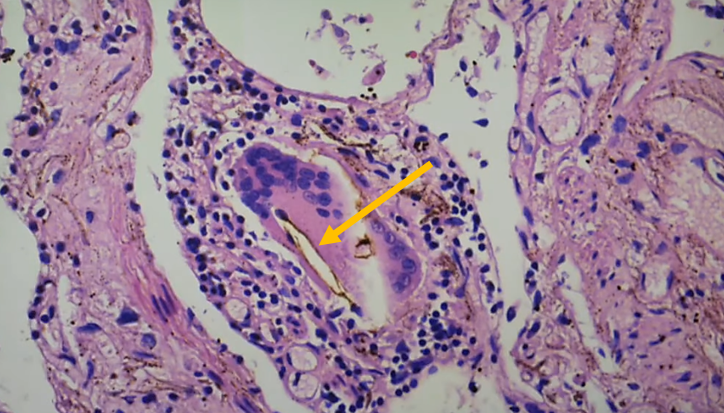
(22/n) Highly inflamed lung tissue with a foreign body. Image
(23/n) Dark field microscopy image of the tissue with the foreign body. Is the foreign body from the vaccine? Image
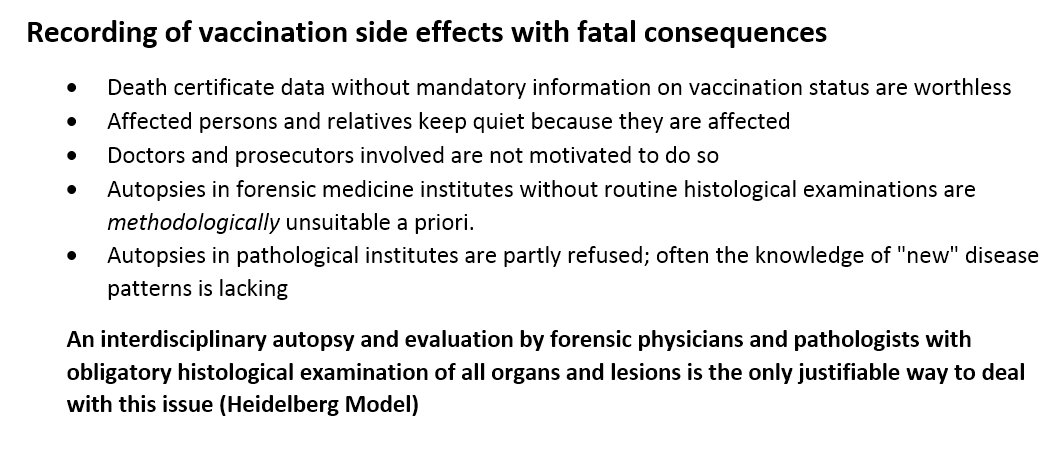
(24/n) There is a problem with recording lethal vaccination side effects: Image

• • •

Missing some Tweet in this thread? You can try to force a refresh
 

Keep Current with Dr John B.

Dr John B. Profile picture

Stay in touch and get notified when new unrolls are available from this author!

Read all threads

This Thread may be Removed Anytime!

PDF

Twitter may remove this content at anytime! Save it as PDF for later use!

Try unrolling a thread yourself!

how to unroll video
  1. Follow @ThreadReaderApp to mention us!

  2. From a Twitter thread mention us with a keyword "unroll"
@threadreaderapp unroll

Practice here first or read more on our help page!

More from @DrJohnB2

Jun 29
(1/n)
What are these generally white blood clots found by various embalmers, probably mainly related to COVID-19 vaccinations?
Embalmer Richard Hirschman (@r_hirschman) has now published some interesting findings about these clots ().
Here is a summary and discussion.
These are the proteins that were found:Image
(2/n)
The most abundant protein is fibrinogen (fibrinogen chains alpha, beta & gamma) (making 56% of total clot protein abundance). Followed by hemoglobin (subunit beta and alpha).
Fibronectin is an important protein for cell adhesion (and in a clot is normally covalently cross-linked to fibin by Factor XIII).
Fibrinogen chains are the main constituents of blood clots, and the presence of relatively large amounts of fibronectin is typical. See, for example, the study of plasma clots by Michał Ząbczyk et al. [1]:

[1] sciencedirect.com/science/articl…Image
(3/n)
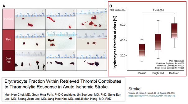
The hemoglobin proteins (beta & alpha subunits) indicate the presence of erythrocytes or the hemoglobin of the erythrocytes (free hemoglobin, Hb).
Hb is known to bind to von Willebrand Factor (VWF), inducing the formation of platelet aggregates on fibrinogen-rich surfaces [1]. The amount of erythrocytes/Hb present in clots varies. Choi et al [2] showed that the more erythrocytes there are in a clot, the more red it is:

[1]
[2] ashpublications.org/blood/article/…
ahajournals.org/doi/full/10.11…Image
Read 16 tweets
Oct 10, 2023
(1/7)
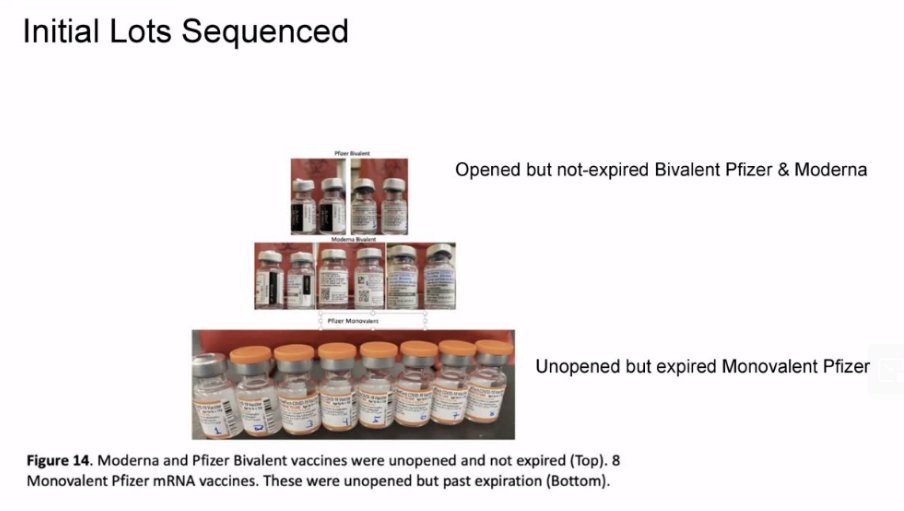
"Plasmid derived dsDNA contamination in mRNA vaccines"
Important lecture from yesterday by Dr. Kevin McKernan (@Kevin_McKernan).
Video:
Slides: worldcouncilforhealth.org/newsroom/
Image
(2/7)


Image
Image
Image
Image
(3/7)


Image
Image
Image
Image
Read 7 tweets
May 4, 2023
(1/11)
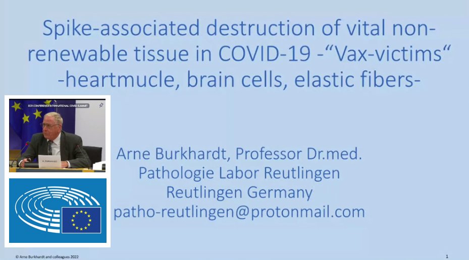
Important presentation by pathologist Prof. Dr. Arne Burkhardt (Germany) yesterday (3 May 2023) in the EU Parliament at the International COVID Summit III on his discoveries in 75 autopsies of people who died in connection with COVID-19 vaccination.
Summary of his slides: Image
(2/11)
- In 77% of the autopsies, the findings indicate that «vaccination had an important impact on the death process»
- «... all organs are affected»
- «... the spike protein is in practially in all organs, mainly in the vessels»
- Image of vessel damage: Image
(3/11)
- Heart tissue with desintegrated musscle fibers and lymphocytic infiltration
- Detection of spike protein in the heart tissue Image
Read 11 tweets
Nov 27, 2022
(1/9) The blood clot mystery
The new documentary “Died Suddenly” (rumble.com/v1wac7i-world-…) shows impressive footage of unusual blood cloths (microclots & white fibrous clots) of deceased vaccinated people.
Here is a compilation of the images: Image
(2/9) Image
(3/9) Image
Read 9 tweets
May 3, 2022
(1/n) Important new discovery:
Not only viruses can be transmitted by air but also ANTIBODIES!
Aerosol-mediated antibody transfer.
" ... an entirely unrecognized mechanism ..."
medrxiv.org/content/10.110… Image
(2/n) Anti-SARS-CoV-2 specific antibodies were found in the eluted from surgical face masks worn by vaccinated lab members donated at the end of one workday: Image
(3/n) With regard to the #CovidVaccine, UNVACCINATED children of VACCINATED adults were found to have anti-SARS-CoV-2 specific antibodies! Children of UNVACCINATED adults did not have them.
Parents who got the #CovidVaccine seem to transmit antibodies in the exhaled aerosol. Image
Read 4 tweets
Jan 15, 2022
(1/6) Interesting & important data: #COVID19 cases, hospitalizations & deaths as a function of time from 1st & 2nd #Covidvaccine dose.
Data from Alberta, Canada.
Unfortunately deleted from the website.
Archived: web.archive.org/web/2022011400… Image
(2/6) COVID-19 CASES after 1st and 2nd Covid vaccine dose.
A large peak in the period of about 0-14 days after the first vaccination and after about 6 months after the second vaccination. Image
(3/6) COVID-19 HOSPITALIZATIONS after 1st and 2nd Covid vaccine dose.
The same pattern. Image
Read 6 tweets

Did Thread Reader help you today?

Support us! We are indie developers!


This site is made by just two indie developers on a laptop doing marketing, support and development! Read more about the story.

Become a Premium Member ($3/month or $30/year) and get exclusive features!

Become Premium

Don't want to be a Premium member but still want to support us?

Make a small donation by buying us coffee ($5) or help with server cost ($10)

Donate via Paypal

Or Donate anonymously using crypto!

Ethereum

0xfe58350B80634f60Fa6Dc149a72b4DFbc17D341E copy

Bitcoin

3ATGMxNzCUFzxpMCHL5sWSt4DVtS8UqXpi copy

Thank you for your support!

Follow Us!

:(