
GI System Histology: Tweetorial #3
Topic: Histology of the Stomach
Pop Quiz (poll in next tweet): How many layers of smooth muscle are present in the Muscularis Externa layer of the stomach?
#HistoHQ
1/16
Topic: Histology of the Stomach
Pop Quiz (poll in next tweet): How many layers of smooth muscle are present in the Muscularis Externa layer of the stomach?
#HistoHQ
1/16

How many layers are found in the stomach Muscularis Externa?
2/16
2/16
Today, we’ll focus our attention on the stomach! Similar to the esophagus (and the rest of the alimentary canal), the stomach possesses the four characteristic layers of the GI tract:
(1) Mucosa
(2) Submucosa
(3) Muscularis Externa
(4) Serosa/Adventitia
3/16
(1) Mucosa
(2) Submucosa
(3) Muscularis Externa
(4) Serosa/Adventitia
3/16

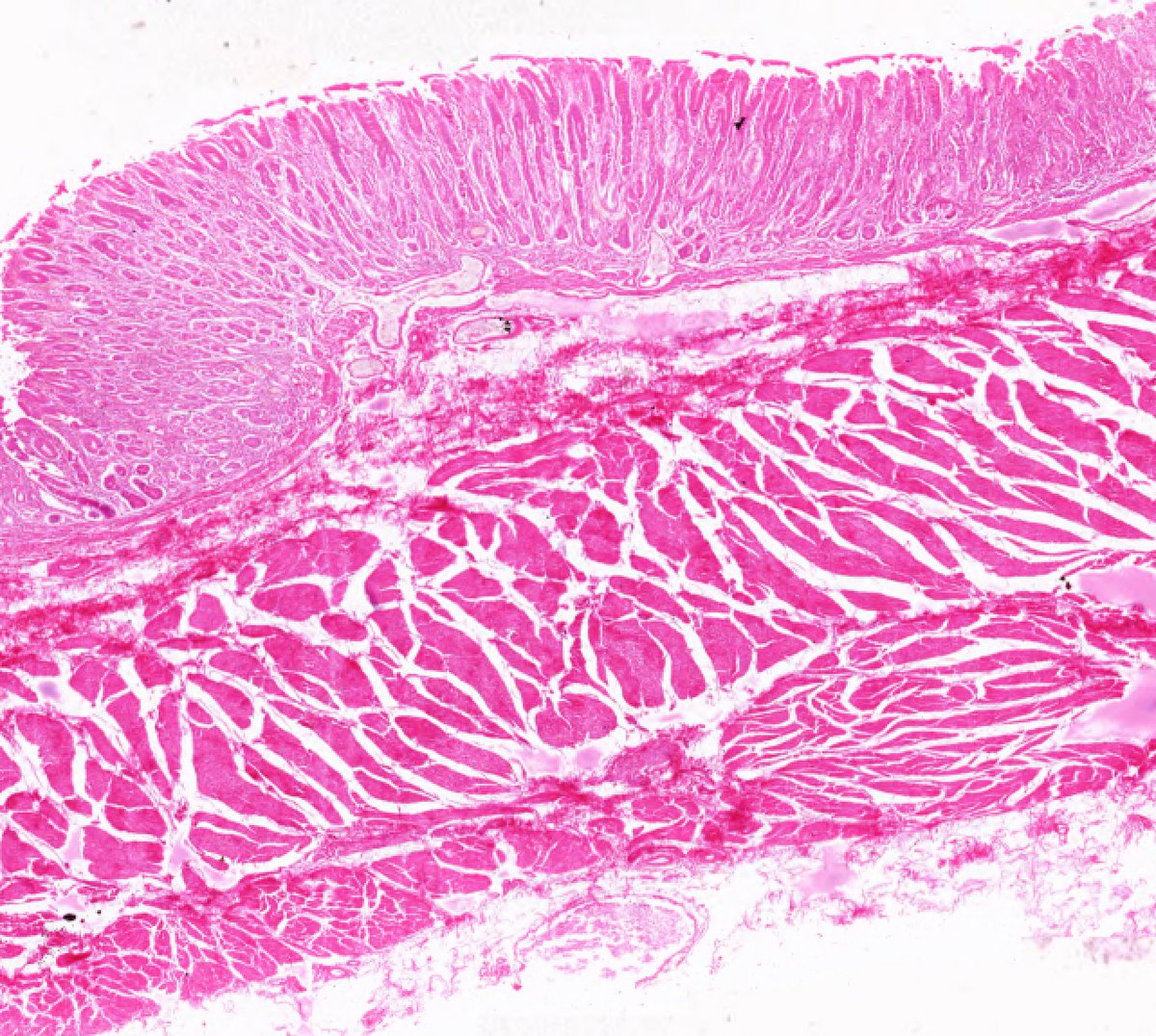
The stomach has some general histologic trends!
Within the mucosa, simple columnar-lined gastric pits lead into gastric glands and occupy much of the Lamina Propria.
The Muscularis Externa is composed of 3 layers: Inner Oblique, Middle Circular, and Outer Longitudinal.
4/16
Within the mucosa, simple columnar-lined gastric pits lead into gastric glands and occupy much of the Lamina Propria.
The Muscularis Externa is composed of 3 layers: Inner Oblique, Middle Circular, and Outer Longitudinal.
4/16
The stomach is a complex organ with 4 anatomically distinct regions:
1. Cardia
2. Fundus
3. Body
4. Pylorus
5/16
1. Cardia
2. Fundus
3. Body
4. Pylorus
5/16
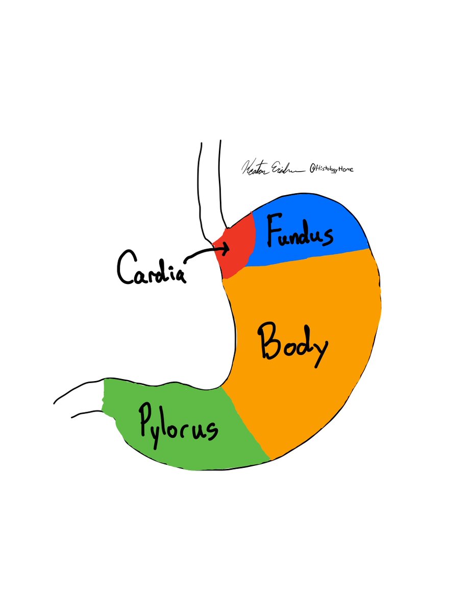
The cardia is a transitional zone between the esophagus and stomach that has a narrow lumen. This portion begins at the Z-line, which marks the change from esophageal stratified squamous to simple columnar epithelium. Its primary function is to produce mucus.
6/16
6/16
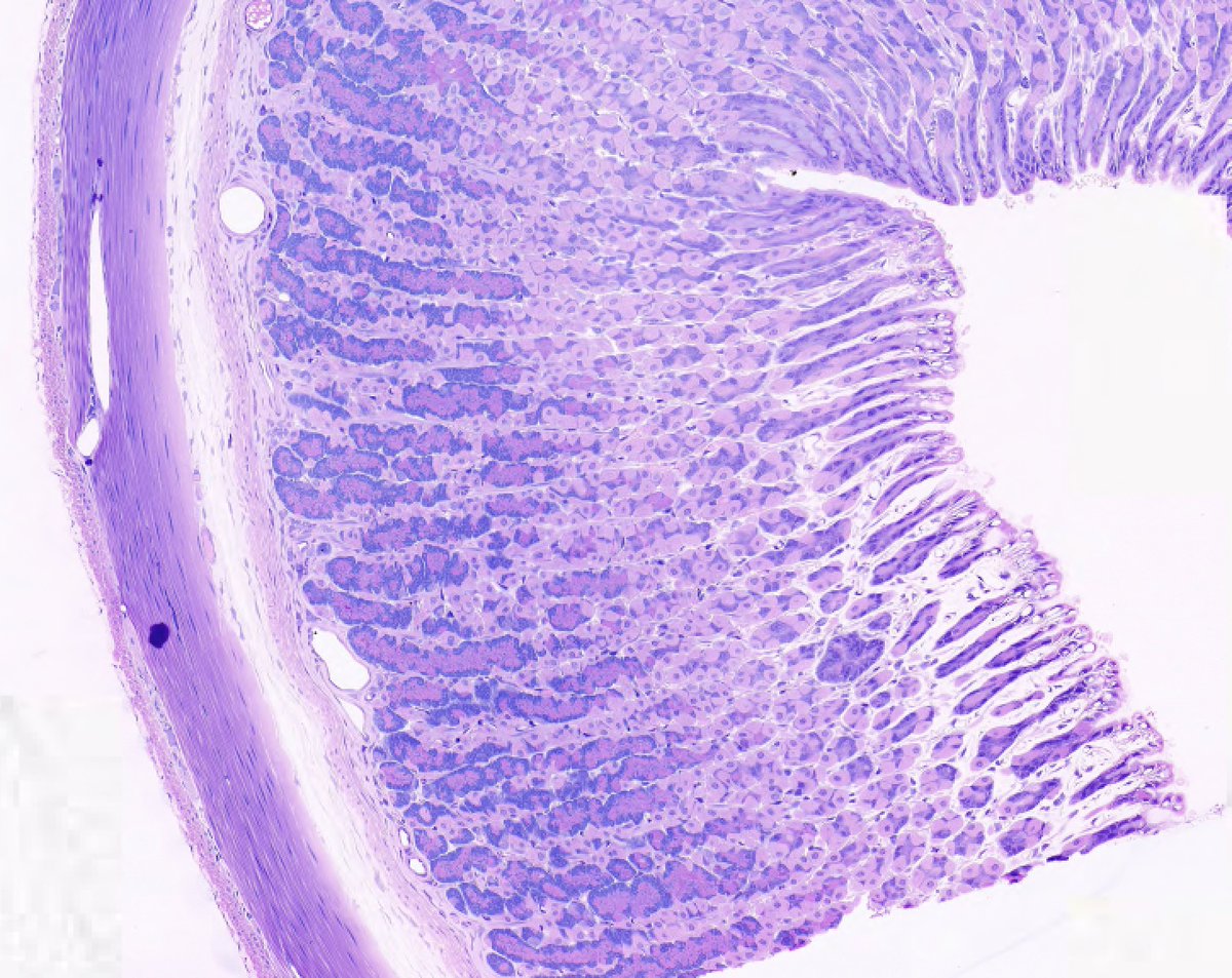
The Fundus and Body are similar histologically! Their functions are to produce gastric acid, pepsinogen, and other secretory products. The glands are very pronounced and developed in these sections!
7/16
7/16
Finally, the pylorus is very similar histologically to the cardia because its function is identical: to primarily produce mucus. This is a narrowing of the stomach immediately proximal to the pyloric sphincter.
8/16
8/16
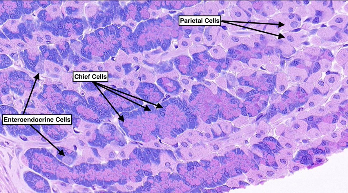
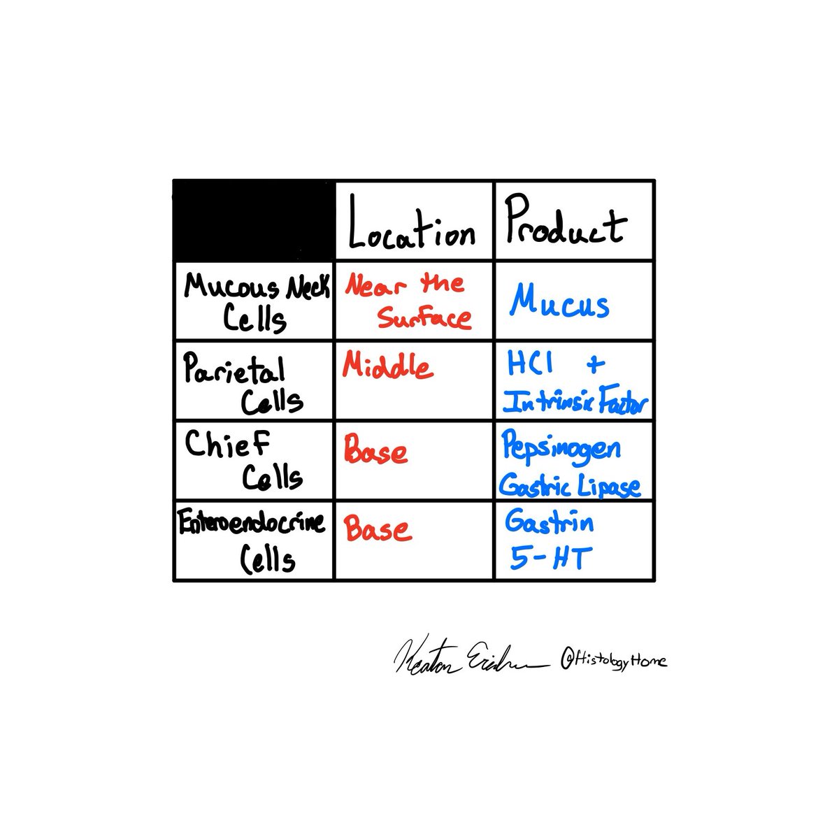
Now, let’s take some time to hone in on the gastric glands!
There are 4 main cell types:
1. Mucous Neck Cells - Not pictured here
2. Parietal Cells
3. Chief Cells
4. Enteroendocrine Cells
9/16
There are 4 main cell types:
1. Mucous Neck Cells - Not pictured here
2. Parietal Cells
3. Chief Cells
4. Enteroendocrine Cells
9/16
This table summarizes the secretory products and relative locations of the cells!
10/16
10/16
High yield points to remember about the stomach:
1. 3 layers of smooth muscle in the Muscularis Externa.
2. Cardia and Pylorus are primarily concerned with mucus production.
3. Body and Fundus are primarily concerned with gastric acid and pepsinogen.
11/16
1. 3 layers of smooth muscle in the Muscularis Externa.
2. Cardia and Pylorus are primarily concerned with mucus production.
3. Body and Fundus are primarily concerned with gastric acid and pepsinogen.
11/16
Let’s test what we’ve learned! Where are we located within the stomach with this section?
12/16
12/16
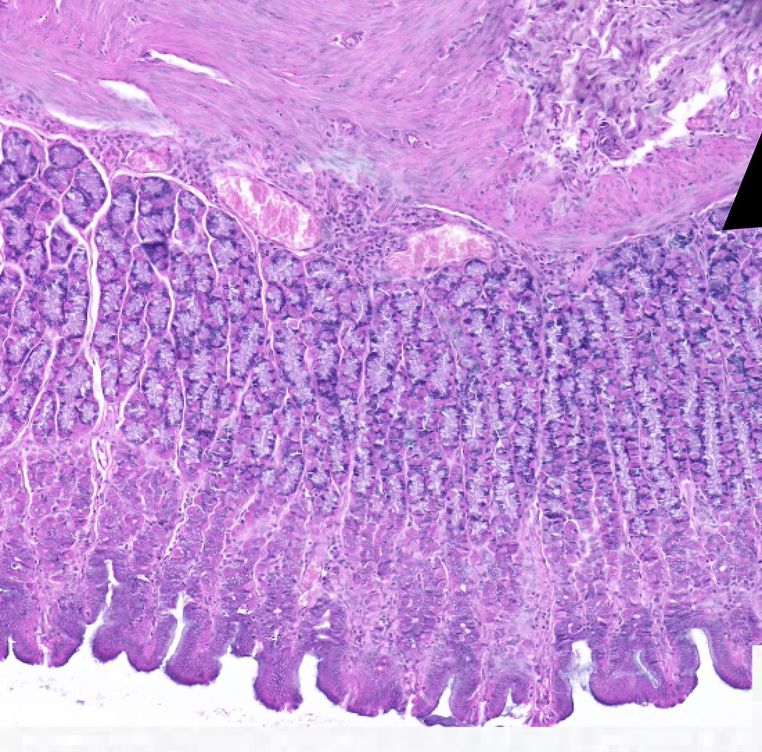
What region of the stomach is shown in the image in the previous tweet?
13/16
13/16
One more test! What cell type is responsible for the production of pepsinogen within the gastric glands?
14/16
14/16
Thanks for sticking around for another week! This week’s #PathCorrelate is gastric ulcers. This is the pathological, focal loss of the mucosal layer of the stomach.
15/16
15/16
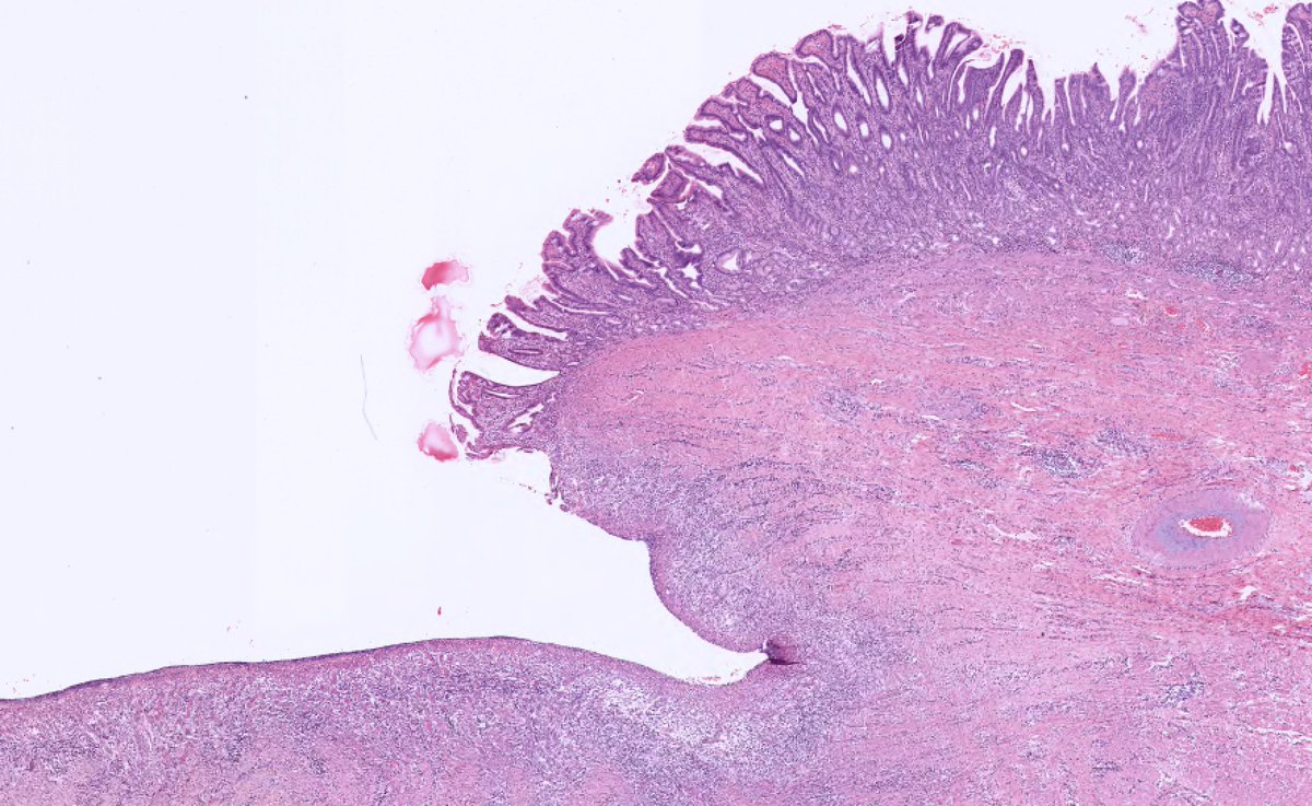
A common presenting symptom is pain after meals. Risk factors include Helicobacter Pylori infection, excessive/prolonged NSAID use, and an imbalance of the local acidity within the stomach lumen.
#PathCorrelate
16/16
#PathCorrelate
16/16
@KMirza @cullen_lilley @CArnold_GI @Dr_Brian_Cox @IsabellaDishong @instapathbio @IR_AMauntie @DeanEhrlich_MD @bgcampmph @tracycookFP @JaredAhrendsen @NicoleJacksonMD @PocketPathCP @BSharpeNotFlat @sarahjlewis9 @Sara_Jiang @ALBoothMD @siparksmd @Path_SIG @jasonaskvig
• • •
Missing some Tweet in this thread? You can try to
force a refresh


