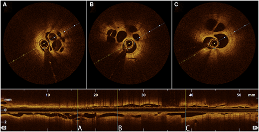
1/24 This month’s #EHJCaseReports #tweetorial focuses on percutaneous left atrial appendage occlusion.
Is #LAAO the exact solution for preventing stroke & embolism in #AF pts who can’t tolerate #OAC?
Or, a new problem while seeking a solution?
doi.org/10.1093/ehjcr/…
#CardioEd
Is #LAAO the exact solution for preventing stroke & embolism in #AF pts who can’t tolerate #OAC?
Or, a new problem while seeking a solution?
doi.org/10.1093/ehjcr/…
#CardioEd
2/24
🎯 The rationale of #LAAO
🔸 In non-valvular #AF patients, 90% of thrombus is located in the #LAA
🔸 These thrombi embolise to the 🧠 & cause #stroke
🎯 Don't miss the ‘#EHRA_ESC #EAPCI expert consensus document on catheter-based #LAAO –an update’ doi.org/10.1093/europa…
🎯 The rationale of #LAAO
🔸 In non-valvular #AF patients, 90% of thrombus is located in the #LAA
🔸 These thrombi embolise to the 🧠 & cause #stroke
🎯 Don't miss the ‘#EHRA_ESC #EAPCI expert consensus document on catheter-based #LAAO –an update’ doi.org/10.1093/europa…

3/24
#LAA closure devices should be safe, efficent and easy to deploy.
📌 Currently available catheter-based devices for #LAAO have different principles.
A- Plug ➡️ Watchman
B- Pacifier ➡️ Amplatzer Amulet
C- Ligation ➡️ Lariat
doi.org/10.1093/europa…
#LAA closure devices should be safe, efficent and easy to deploy.
📌 Currently available catheter-based devices for #LAAO have different principles.
A- Plug ➡️ Watchman
B- Pacifier ➡️ Amplatzer Amulet
C- Ligation ➡️ Lariat
doi.org/10.1093/europa…

4/24
#LAA has different morphologies based on Wang’s classification.
Cactus (∼30%)
Chicken-wing (∼48%)
Windsock (∼19%)
Cauliflower (∼3%)
journals.plos.org/plosone/articl…
#LAA has different morphologies based on Wang’s classification.
Cactus (∼30%)
Chicken-wing (∼48%)
Windsock (∼19%)
Cauliflower (∼3%)
journals.plos.org/plosone/articl…
5/24
🎯 Pre- & peri- & post-procedural imaging w/ #TEE and #yesCT is key for:
👉 Ruling out #LAA thrombus
👉 #LAA anatomy & dimensions
👉 Equipment (device) selection
👉 Guiding procedural device implantation
👉 Device surveillance post-implantation
doi.org/10.1093/europa…
🎯 Pre- & peri- & post-procedural imaging w/ #TEE and #yesCT is key for:
👉 Ruling out #LAA thrombus
👉 #LAA anatomy & dimensions
👉 Equipment (device) selection
👉 Guiding procedural device implantation
👉 Device surveillance post-implantation
doi.org/10.1093/europa…

6/24 Persistent large (≥5 mm) peri-device leaks detected w/ post-procedural imaging deserve long-term #OAC or a second occlusion attempt.
🎯 As an alternative strategy, this case shows successful percutaneous leak closure w/ an Amplatzer Vascular Plug II doi.org/10.1093/ehjcr/…
🎯 As an alternative strategy, this case shows successful percutaneous leak closure w/ an Amplatzer Vascular Plug II doi.org/10.1093/ehjcr/…
7/24
📌 Imaging w/ #TEE is essential during transseptal puncture.
📌 For most #LAA, where the body is superior-anterior directed, a standard infer-posterior puncture of the fossa ovalis is recommended to provide a direct vector towards the superior-anterior LAA.
📌 Imaging w/ #TEE is essential during transseptal puncture.
📌 For most #LAA, where the body is superior-anterior directed, a standard infer-posterior puncture of the fossa ovalis is recommended to provide a direct vector towards the superior-anterior LAA.

8/24
#LAAO is still possibile in patients w/ a previous atrial septal device.
📌 In this case report you will find out how #LAAO was performed w/ a LAmbre device in a patient w/ previously implanted #PFO occluder. doi.org/10.1093/ehjcr/…

#LAAO is still possibile in patients w/ a previous atrial septal device.
📌 In this case report you will find out how #LAAO was performed w/ a LAmbre device in a patient w/ previously implanted #PFO occluder. doi.org/10.1093/ehjcr/…


9/24
📌 Pericardial tamponade rates during the #LAAO is ~ 1 %.
👉 This case shows cardiac tamponade due to high PSI injection during #LAA angiography and sealing the perforation w/ Watchman device.
doi.org/10.1093/ehjcr/…
📌 Pericardial tamponade rates during the #LAAO is ~ 1 %.
👉 This case shows cardiac tamponade due to high PSI injection during #LAA angiography and sealing the perforation w/ Watchman device.
doi.org/10.1093/ehjcr/…

10/24
#yesCT #TEE & fluoroscopic measurements for the appropriate size of Amulet device:
🎯 #LAA orifice ➡️ yellow lines
🎯 Landing zone ➡️ 10-12 mm inside the orifice (red lines)
🔑 For appropriate measurements mean left atrial pressure should be >12 mmHg
#yesCT #TEE & fluoroscopic measurements for the appropriate size of Amulet device:
🎯 #LAA orifice ➡️ yellow lines
🎯 Landing zone ➡️ 10-12 mm inside the orifice (red lines)
🔑 For appropriate measurements mean left atrial pressure should be >12 mmHg

11/24
🔑 The appropriate size of the Amulet device is decided according to the ‘maximum landing zone’, which is 10-12 mm from the ostium of the #LAA.
🔑 For an #LAA w/ a landing zone of 16 mm 👉 20 mm Amulet device is recommended.
🔑 The appropriate size of the Amulet device is decided according to the ‘maximum landing zone’, which is 10-12 mm from the ostium of the #LAA.
🔑 For an #LAA w/ a landing zone of 16 mm 👉 20 mm Amulet device is recommended.

12/24
#LAAO is performed under #TEE & fluoroscopic guidance.
🎯 RAO 30/cranial 20 👉 proximal #LAA
🎯 RAO 30/caudal 20 👉 distal #LAA
🔑🕵️♂️
RAO 30/cranial 20 ↩️ #TEE 45
RAO 30/caudal 20 ↩️ #TEE 135
eurointervention.pcronline.com/article/left-a…

#LAAO is performed under #TEE & fluoroscopic guidance.
🎯 RAO 30/cranial 20 👉 proximal #LAA
🎯 RAO 30/caudal 20 👉 distal #LAA
🔑🕵️♂️
RAO 30/cranial 20 ↩️ #TEE 45
RAO 30/caudal 20 ↩️ #TEE 135
eurointervention.pcronline.com/article/left-a…


13/24
📌Amulet device deployment has 4️⃣ steps:
1 > Unsheat for ‘ball’ configuration
2-3 > Advance delivery cable for ‘triangle’ configuration and ‘lobe’ unfolded
4 > Unsheat for ‘Disc’ unfolded
🎯 Lobe of the device should sit landing zone.
doi.org/10.1093/europa…
📌Amulet device deployment has 4️⃣ steps:
1 > Unsheat for ‘ball’ configuration
2-3 > Advance delivery cable for ‘triangle’ configuration and ‘lobe’ unfolded
4 > Unsheat for ‘Disc’ unfolded
🎯 Lobe of the device should sit landing zone.
doi.org/10.1093/europa…

14/24
📌 Please find step by step Amulet device deployment + tug test in the ⬇️ videos.
1st video ➡️ ‘ball’ & ‘triangle’ configuration of the ‘lobe’
2nd video ➡️ ‘disc’ deployment
3rd video ➡️ tug test
doi.org/10.1093/europa…
📌 Please find step by step Amulet device deployment + tug test in the ⬇️ videos.
1st video ➡️ ‘ball’ & ‘triangle’ configuration of the ‘lobe’
2nd video ➡️ ‘disc’ deployment
3rd video ➡️ tug test
doi.org/10.1093/europa…
15/24
🎯 Before release, 5️⃣ signs of device stability need to be verified.
1️⃣ Slightly compressed ‘lobe’
2️⃣ Orientation between the ‘lobe’ and #LAA orifice
3️⃣ Concave shaped ‘disc’
4️⃣ Separated ‘disc’ & ‘lobe’
5️⃣ The midpoint of the ‘lobe’ should be distal to the LCx.
🎯 Before release, 5️⃣ signs of device stability need to be verified.
1️⃣ Slightly compressed ‘lobe’
2️⃣ Orientation between the ‘lobe’ and #LAA orifice
3️⃣ Concave shaped ‘disc’
4️⃣ Separated ‘disc’ & ‘lobe’
5️⃣ The midpoint of the ‘lobe’ should be distal to the LCx.

16/24
⁉️ Despite all efforts, embolisation of the device can still occur.
🎯 In this case report, the embolised #LAAO device was retrieved using a double snaring technique.
doi.org/10.1093/ehjcr/…
⁉️ Despite all efforts, embolisation of the device can still occur.
🎯 In this case report, the embolised #LAAO device was retrieved using a double snaring technique.
doi.org/10.1093/ehjcr/…
17/24
#LAAO to who?
🎯 High-risk non-valvular #AF patients who ‘ve contraindications or can't tolerate long-term #OAC.
🎯 Patients w/elevated bleeding risk
🎯 Patients suffering from stroke while on #OAC
🎯 Patients unwilling to receive #OAC
doi.org/10.1093/europa…

#LAAO to who?
🎯 High-risk non-valvular #AF patients who ‘ve contraindications or can't tolerate long-term #OAC.
🎯 Patients w/elevated bleeding risk
🎯 Patients suffering from stroke while on #OAC
🎯 Patients unwilling to receive #OAC
doi.org/10.1093/europa…


18/24
⁉️ Optimal post #LAAO antithrombotic 💊 & duration remain controversial.
The results of ongoing
📌 ANDES
📌 ADRIFT
📌 SAFE-LAAC
trials comparing different post #LAAO antithrombotic regimens will shed light on this issue.
doi.org/10.1093/europa…
⁉️ Optimal post #LAAO antithrombotic 💊 & duration remain controversial.
The results of ongoing
📌 ANDES
📌 ADRIFT
📌 SAFE-LAAC
trials comparing different post #LAAO antithrombotic regimens will shed light on this issue.
doi.org/10.1093/europa…

19/24
⁉️ Device-related thrombus (3.75%) is related to concurrent #OAC or #DAPT 💊 and is associated w/ ⬆️ risk of ischemic stroke.
👉 See the following case report for how device-related thrombus was managed in an 85 yo 👴 w/ a history of #LAAO
doi.org/10.1093/eurhea…
⁉️ Device-related thrombus (3.75%) is related to concurrent #OAC or #DAPT 💊 and is associated w/ ⬆️ risk of ischemic stroke.
👉 See the following case report for how device-related thrombus was managed in an 85 yo 👴 w/ a history of #LAAO
doi.org/10.1093/eurhea…
20/24
⁉️Another question to be answered:
🤔 Is #LAAO better than, or at least as safe and effective as antithrombotic agents?
Without a doubt, ongoing studies
📌 OCCLUSION-AF
📌 STROKE-CLOSE
📌 CLOSURE-AF
📌 ASAP-TOO
will provide essential insights.
doi.org/10.1093/europa…
⁉️Another question to be answered:
🤔 Is #LAAO better than, or at least as safe and effective as antithrombotic agents?
Without a doubt, ongoing studies
📌 OCCLUSION-AF
📌 STROKE-CLOSE
📌 CLOSURE-AF
📌 ASAP-TOO
will provide essential insights.
doi.org/10.1093/europa…

21/24
🎯 The results of PRAGUE-17 trial, which compares NOACs w/ #LAAO in high-risk #AF patients (CHA2DS2-VASC ≥3), showed #LAAO was non-inferior to NOACs in preventing major AF-related cardiovascular, neurological, and bleeding events.
eurointervention.pcronline.com/trials/left-at…
🎯 The results of PRAGUE-17 trial, which compares NOACs w/ #LAAO in high-risk #AF patients (CHA2DS2-VASC ≥3), showed #LAAO was non-inferior to NOACs in preventing major AF-related cardiovascular, neurological, and bleeding events.
eurointervention.pcronline.com/trials/left-at…

22/24
⁉️ Which device is better ⁉️
🎯 AMULET-IDE study (Hot Line #ESCCongress2021) compared Amulet vs WATCHMAN device in high-risk #AF patients.
🎯 The Amulet device was associated w/
⬆️ #LAA occlusion
↔️ Safe & effective for stroke prev.
compared to the Watchman device.
⁉️ Which device is better ⁉️
🎯 AMULET-IDE study (Hot Line #ESCCongress2021) compared Amulet vs WATCHMAN device in high-risk #AF patients.
🎯 The Amulet device was associated w/
⬆️ #LAA occlusion
↔️ Safe & effective for stroke prev.
compared to the Watchman device.

23/24
🤔 Do we have alternative venous access sites for #LAAO ?
🙂 The answer is YES.
🎯 See the ⬇️ case report, which demonstrates transhepatic access in a patient w/ poor femoral access for implantation of the #LAAO device.
doi.org/10.1093/ehjcr/…
🤔 Do we have alternative venous access sites for #LAAO ?
🙂 The answer is YES.
🎯 See the ⬇️ case report, which demonstrates transhepatic access in a patient w/ poor femoral access for implantation of the #LAAO device.
doi.org/10.1093/ehjcr/…

24/24
We hope you enjoyed our #tweetorial on #LAAO
Let's say goodbye with a poll.
⁉️ Which #LAAO device has the ‘8’ image on #TTE ?
⬇️ Please vote ⬇️
Thank you to our fabulous Twitter Editors: @aayshacader @FarhanaAra @KardiologieHH @TJ_Yeo @ANazmiCalik
#EHJCaseReports
We hope you enjoyed our #tweetorial on #LAAO
Let's say goodbye with a poll.
⁉️ Which #LAAO device has the ‘8’ image on #TTE ?
⬇️ Please vote ⬇️
Thank you to our fabulous Twitter Editors: @aayshacader @FarhanaAra @KardiologieHH @TJ_Yeo @ANazmiCalik
#EHJCaseReports

• • •
Missing some Tweet in this thread? You can try to
force a refresh


