“COVID toes” are swollen discolored toes (and fingers) that were seen in areas with high incidence of COVID-19, but the cause is unknown. This new study by @JeffGehlhausen et al shows lack of association between covid toes and SARS-CoV-2 infection. 🧵(1/)
We enrolled 23 pandemic chilblains (PC) patients. While there is an association with community COVID cases (blue line) and PC (red bars), only 2 PC patients had evidence of infection by PCR or antibodies. We wondered if people may have missed the time window for testing +ve. (2/)
PCR testing was difficult to access at the time of initial wave (2020). Thus, we employed two distinct measures of antibodies - ELISA and @serimmune SERA assays - against SARS-CoV-2 S, RBD and N. Only 2 of the 23 patients (who were also PCR +ve) had consistent antiviral Abs. (3/)
However, Ab responses wane over time. So we next tested TCR repertoire of the PC patients using @AdaptiveBiotech’s immunoSEQ T-MAP COVID platform. This assay is more sensitive than Ab assay. It picked up one additional patient, PC #10, as testing pos for antiviral T cells. (4/)
Using an orthogonal approach, we conducted a T cell stimulation assays of PBMCs utilizing a pool of S-protein peptides. This analysis also picked up patient #10 as being positive for T cells against S (consistent with immunoSEQ data). Thus patient #10, 12 and 16 are positive.(5/)
Spike antigen in the toe has been detected by some and suggested as a cause of PC.When we used anti-S Ab for immunohistochemistry (IHC), we saw nice signals for spike in the PC skin (left). However, the same Ab also stained pre-pandemic tissues (right) -> Ab non-specific. (6/)
In fact, we tested a few commercially available antibodies to the S and N and found cross-reactivity to human tissues from the prepandemic era. This is why we need multiple different approaches to ensure what we see in IHC is real. (7/)
So only 3 of 23 pandemic chilblains patients were found to be COVID positive by PCR/Ab/T tests. What accounts for the rest of the patients’ skin lesions? We tested autoantibodies to human exoproteome with REAP with @Aaronmring team and found no increase in PC over control. (8/)
So what can explain pandemic chilblains? Strong mucosal innate immune responses (interferons) that eliminated virus at exposure but caused delayed onset skin inflammation? Seronegative abortive infections by cross-protective T-cells that somehow led to delayed skin rash? (9/)
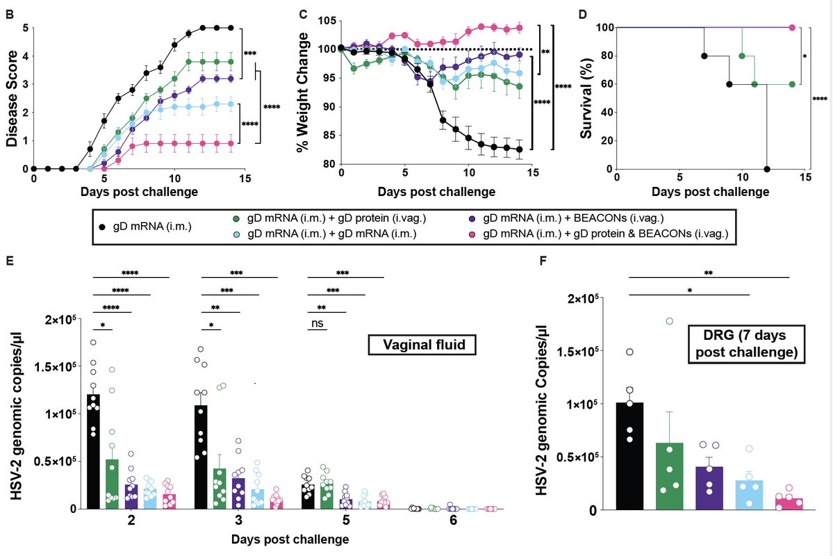
Introducing BEACON (Bioactive Enhanced Adjuvant Chemokine Oligonucleotide Nanoparticles) to stimulate mucosal immune responses against genital #HSV infection. Awesome work led by @sachinbhag, who designed and developed BEACON to guide T and B cells (1/) biorxiv.org/content/10.110…
The BEACON nanoparticle is composed of CpG ODN (TLR9 agonist) and CXCL9 (chemokine). When applied to the vaginal mucosa, the recruitment of antiviral T cells is achieved with minimal local inflammation - much more potent than CpG or CXCL9 separately. (2/)
Intramuscular vaccines do not promote mucosal immunity. BEACON can be used as a local adjuvant to boost HSV-2 gD- or gB-specific T and B cells, preventing not only disease/death but also blocking viral load in both vaginal tissue and dorsal root ganglia (🚫latency)(3/)👇🏼
A fascinating new study by Vishnu Shankar et al. @stanfordimmuno shows that oxidative stress is a shared characteristic of ME/CFS and Long COVID in lymphocytes due to inability to clear reactive oxygen species. This happens in sex-specific manner. (1/) pnas.org/doi/abs/10.107…
Females show higher mtROS levels and insufficient antioxidant levels, while males show mitochondrial lipid oxidative damage. While the reason for this is unclear, it may explain the sex differences in lymphocyte dysfunction we see in PAIS in general. (2/) science.org/doi/10.1126/sc…
ROS-targeting therapies were tested. Metformin treatment in vitro showed some impact on CD4 T cell proliferation. I suspect that other therapies to induce autophagy/mitophagy might also benefit restoration of T cell phenotype. #LowDoseRapamycin 👇🏼 (3/) polybio.org/projects/long-…
Published today! Victoria Bastos, @KerrieGreene_ et al found two distinct immunotypes of ME/CFS based on the cerebrospinal fluid analysis. Great collaboration with @MBVanElzakker @microbeminded2 and the Bragée clinic in Sweden. (1/) academic.oup.com/jimmunol/artic…
This is perfect timing as Victoria will present these data at the @polybioRF symposium today. (2/)
Based on cerebrospinal fluid cytokines, we identified two clusters of ME/CFS patients. Cluster 1 had elevated matrix metalloproteinases & many cytokines compared to cluster 2. Other than older age (Cluster 1), clinical presentation of these clusters was similar. (3/)
Published today📣
Our nasal booster in the "Prime & Spike" vaccine works without adjuvants (which are needed to induce adaptive immunity but also cause inflammation). @Kwon_Dongil @tianyangmao @BenIsraelow et al. asked how this is possible. (1/) nature.com/articles/s4159…
Prime & Spike is a vaccine strategy that leverages preexisting immunity primed by conventional vaccines to elicit mucosal IgA and T cell responses that prevent COVID infection and transmission in rodents. The nasal booster is simply the spike protein (2/) science.org/doi/10.1126/sc…
Our new study shows that the nasal spike protein booster converts lymph node memory B cells into IgA-secreting cells in the lung with the help of memory CD4 T cells. Ag-specific CD4 T cells replace all the necessary functions of adjuvants without nonspecific inflammation! (3/)
This prospective observational study led by @connorbgrady @bornali_27 @SilvaJ_C @hmkyale examined the impact of the primary COVID-19 vaccination on the symptoms and immune signatures of 16 people with #longCOVID. Here is what we found 👇🏼 (1/)
This study asked: Does COVID vaccination improve symptoms of long COVID? If so, is the improvement due to robust T and B cell responses leading to the clearance of the viral reservoir? If not, is there an immune feature that predicts worsening of LC? (2/)
The self-reported impact of vaccination was variable. Of the 16 long COVID patients, 10 felt better, 3 had no change, and 3 had worse health (1 hospitalized) 12 weeks after vaccination. Both physical and social effects of symptom burden appeared to decrease after vaccination. (3/)
Our preprint on post-vaccination syndrome is out. We studied immune signatures and examined spike protein in the blood of people who have developed chronic illnesses after COVID-19 vaccination. (1/) medrxiv.org/content/10.110…
Vaccines have saved countless lives and inspired me to become an immunologist. While generally safe, some people experience adverse effects, including Post-Vaccination Syndrome (PVS). Studying PVS is crucial for improving patient care and enhancing vaccine safety & acceptance. (2/) pubmed.ncbi.nlm.nih.gov/37986769/