What immune cell features are most predictive of COVID outcomes? @mkuchroo@JcsHuang Patrick Wong et al used ML algorithm Multiscale PHATE to assign each immune cell type in COVID patients a mortality-likelihood score. Latest from @KrishnaswamyLab 💪🏼 (1/) go.nature.com/3K0QCqi
Based on the flow cytometry data on 54 million cells from COVID 168 patients, the low density granulocytes (neutrophils and eosinophils) were the most enriched cell types in patients who had fatal COVID, followed by inflammatory monocytes and certain B cell subsets. (2/)
In contrast, T cells (most of them; see below), NK cells and dendritic cells were associated with the lowest mortality likelihood scores. They are likely protecting the host from lethal disease. (3/)
When you dive into each of the lymphocyte subsets though, things start to look very interesting. Among the CD4 T subsets, while Th1, IL-4+, IL-6+ cells are protective, IL-17* cells that also produce IFN-g & granzyme B (red) have the highest mortality likelihood score. (4/)
Within the CD8 T cell subsets, hyperactivated CD8+ T cell (CD8+CD45RA+TIM3+HLA-DR+PD1+) TEMRA cells expressing granzyme B were correlated with lethality, while naive cells had the lowest mortality likelihood score. (5/)
The beneficial vs. pathogenic roles of T cells in COVID have been noted before. However, the Multiscale PHATE assigned vastly different mortality scores to each T cell subset with distinct effector functions. Seeing this from @mkuchroo was definitely a wow moment for me. (6/)
What about B cells? Plasmablasts (brown) had highest mortality likelihood score. In contrast, a subset of late-activated mature B cells defined by CD86+ (green) was most enriched in patients with good outcomes. Consistent with a previous study. pubmed.ncbi.nlm.nih.gov/32910469/ (7/)
We are so fortunate to work with Dr. @KrishnaswamyLab and her team on this project. Her team keeps innovating new techniques to analyze complex and massive data with method that can learn and visualize cellular features - something immunologists love to do ❤️ (8/)
But they are just getting started 💪🏼 @KrishnaswamyLab also found a way to generate hard to obtain data from easy to obtain ones. For example, they can use ‘feature mapping GAN’ to model patients' flow cytometry data from clinical monitoring data 🤯 (9/)
This collaboration highlights the power of interdisciplinary research. When immunologists work together with computer scientists, amazing insights can emerge (there’s a lot more in this paper). Highlighting all the authors who contributed to the study 👇🏽 (end)
• • •
Missing some Tweet in this thread? You can try to
force a refresh
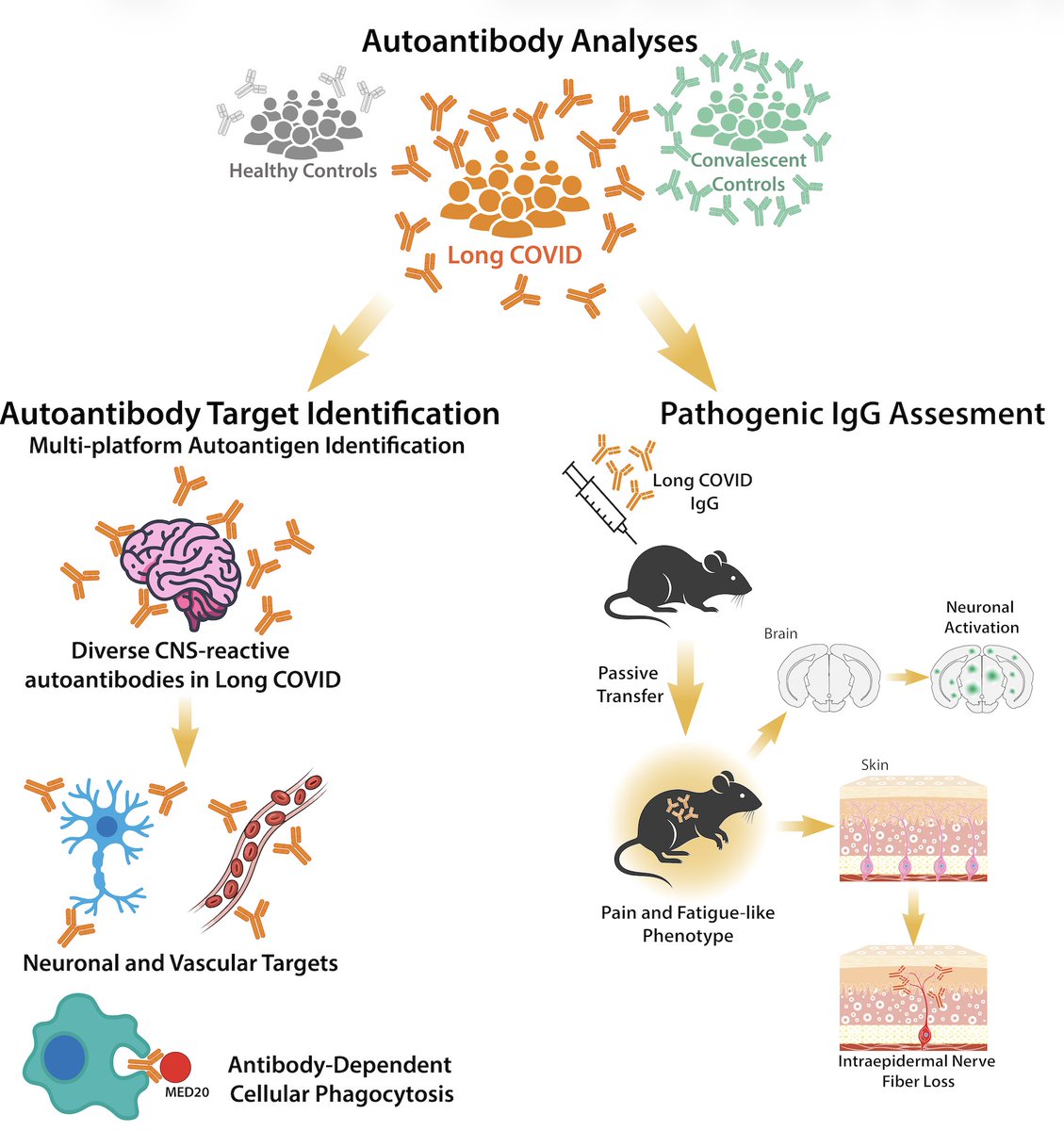
Excited to share our study by @keylas3 et al. on pathological autoantibodies in people with Long COVID. We asked whether IgG in patients with Long COVID bind to human tissues/antigens and cause pathologies when transferred into mice. With @PutrinoLab
Using tissue-based staining, a human proteome array, ELISA, IgG-pull down and mass spec, we identified a wide array of autoantibodies in those with LC. IgG targeting neurological antigens, such as NMDAR, were elevated in LC. Collab with CellTrend and @C_Scheibenbogen 🙏🏼
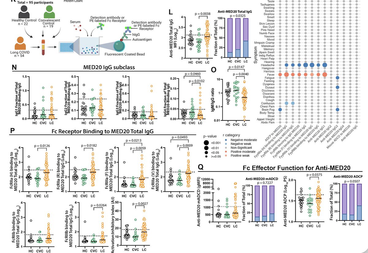
With SeromYx, we found that autoantibodies to MED20 in LC have undergone class switching to IgG from IgM, bind to many FcgRs, and induce robust phagocytosis when in immune complexes compared to IgG from control participants
Is there an association between human herpesviruses (HHVs) reactivation and Long COVID? We analyzed HHV DNA shedding in saliva and found that HHV-6 correlates with Long COVID severity. Claire Laxton, @S_Tabachnikova, Lily Cooke, Kexin Wang et al.
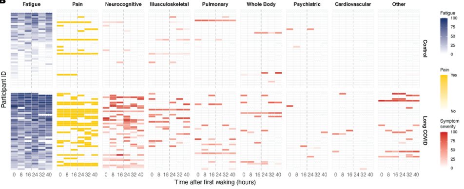
Our study enrolled 45 participants with LC and 45 age-sex-matched controls. Surveys and health questionnaires were used to collect symptom profiles. Note the intense levels of fatigue, pain, and other symptoms in our LC group (bottom half) throughout the days 😱 (2/)
We quantified DNA from multiple HHVs in saliva to ask a simple question: are any of these viruses more active in those with worse Long COVID? HHV-6 stood out. Higher HHV-6 levels were associated with more severe symptoms and greater functional impairment (3/)
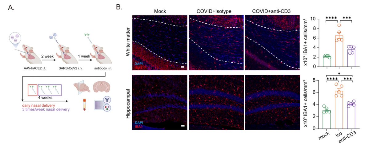
Our new preprint by @peowenlu @SaefIzzy @weinerlabhms and colleagues shows that nasal anti-CD3 monoclonal antibody treatment can reduce neuroinflammation in a mouse model of Long COVID, even when administered at 4 weeks after infection 🧠 biorxiv.org/content/10.648…
Neuroinflammatory damage is a hallmark of Long COVID. Nasal delivery of anti-CD3 mAb induces Treg and has shown therapeutic benefit in various autoimmune and CNS models of disease. In this study, we asked whether anti-CD3 mAb can reduce damage and restore neurogenesis.
First, we tested whether anti-CD3 mAb administered nasally starting at 1 week post-infection for 4 weeks. The treatment restored microglial and astrocyte densities and reduced inflammatory cytokines.
A groundbreaking paper by @younis_sh1 et al. @stanfordimmuno provides an answer to the long-standing question about how EBV infections are linked to lupus. A short thread to explain the key findings. (1/) science.org/doi/10.1126/sc…
The authors developed a new method called EBV-seq, enabling them to overcome the barrier of studying rare cells (~25 per 10,000 B cells in lupus patients) that are infected by EBV. Note that in healthy people, only 1/10,000 B cells are EBV-infected. (2/)
First, the authors found that EBV infects autoreactive B cells that express anti-nuclear antibodies in lupus patients, but not in healthy people or in patients with multiple sclerosis. (3/)
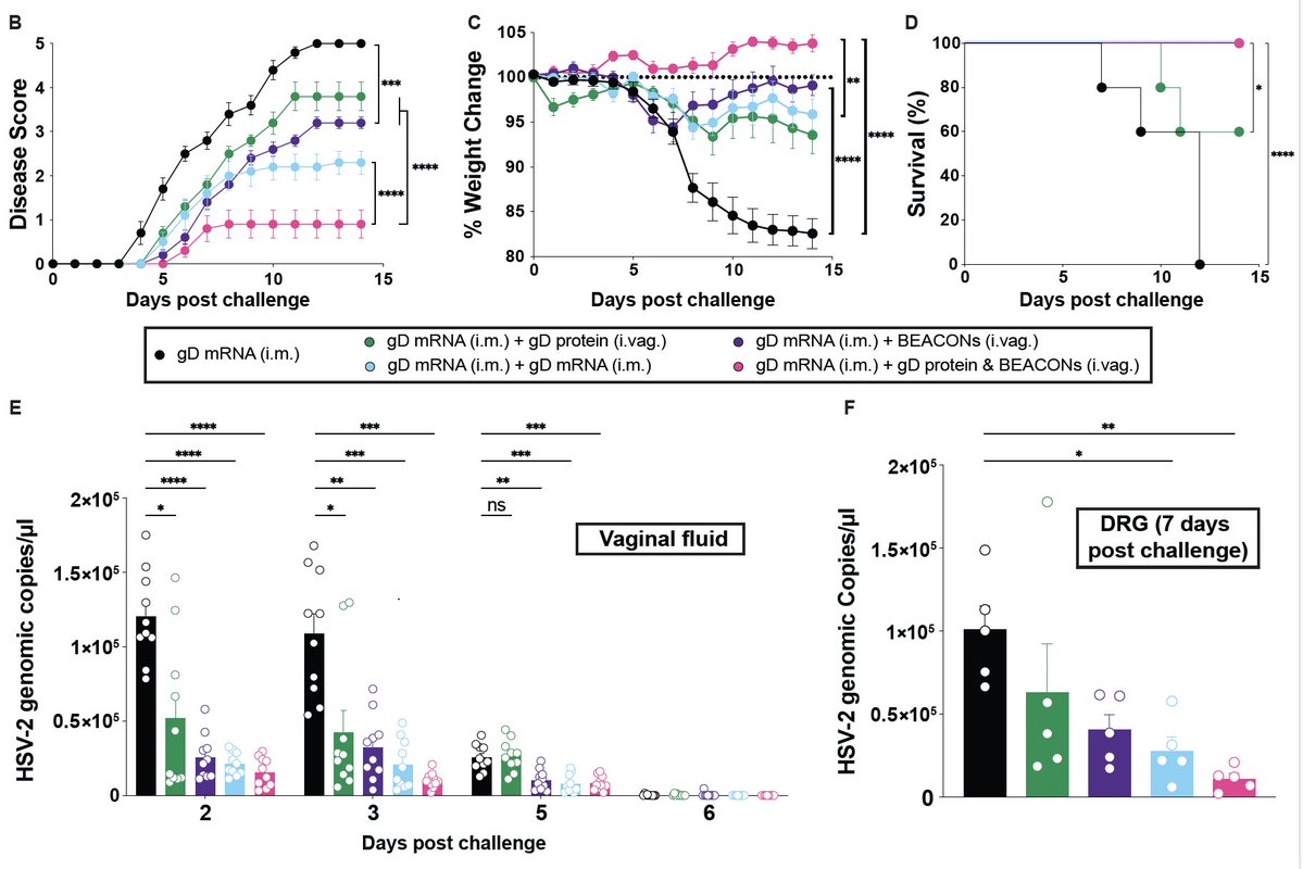
Introducing BEACON (Bioactive Enhanced Adjuvant Chemokine Oligonucleotide Nanoparticles) to stimulate mucosal immune responses against genital #HSV infection. Awesome work led by @sachinbhag, who designed and developed BEACON to guide T and B cells (1/) biorxiv.org/content/10.110…
The BEACON nanoparticle is composed of CpG ODN (TLR9 agonist) and CXCL9 (chemokine). When applied to the vaginal mucosa, the recruitment of antiviral T cells is achieved with minimal local inflammation - much more potent than CpG or CXCL9 separately. (2/)
Intramuscular vaccines do not promote mucosal immunity. BEACON can be used as a local adjuvant to boost HSV-2 gD- or gB-specific T and B cells, preventing not only disease/death but also blocking viral load in both vaginal tissue and dorsal root ganglia (🚫latency)(3/)👇🏼
A fascinating new study by Vishnu Shankar et al. @stanfordimmuno shows that oxidative stress is a shared characteristic of ME/CFS and Long COVID in lymphocytes due to inability to clear reactive oxygen species. This happens in sex-specific manner. (1/) pnas.org/doi/abs/10.107…
Females show higher mtROS levels and insufficient antioxidant levels, while males show mitochondrial lipid oxidative damage. While the reason for this is unclear, it may explain the sex differences in lymphocyte dysfunction we see in PAIS in general. (2/) science.org/doi/10.1126/sc…
ROS-targeting therapies were tested. Metformin treatment in vitro showed some impact on CD4 T cell proliferation. I suspect that other therapies to induce autophagy/mitophagy might also benefit restoration of T cell phenotype. #LowDoseRapamycin 👇🏼 (3/) polybio.org/projects/long-…