Dr. OMID BANDARCHI Profile picture
May 4, 2022 6 tweets 2 min read Read on X
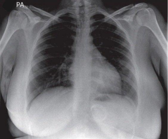
These frontal & lateral views belong to a patient with a history of lung cancer which presented with a newly developed dyspnea. Considering the patient’s history and CXR findings,what is the most likely cause of the elevation of the right hemidiaphragm?
#chestrad #radiology Image
Hint👉 I wanted to discuss about this subject and I incidentally encountered this case! Honestly speaking it's not complicated case ,but the question is worthy of noticing!
So do you think atelectasis of the R.U lobe is the main culprit?The answer based on many references is Not!
🛑Answer part 1
Phrenic nerve paralysis is a common cause of elevation of one side of the diaphragm.
👉Take home message:
The combination of a lung or mediastinal mass and elevation of the diaphragm strongly suggests phrenic nerve paralysis.
Eventration is a different entity!
🛑Answer part 2
Paralysis tion can be confirmed by fluoroscopy, which will reveal paradoxic motion of the diaphragm. A sniff accentuates diaphragmatic motion, so is therefore useful in eliciting paradoxic motion.
Answer part 3
Eventration is similar to paralysis but represents an area of weakness &thinning of the diaphragm. There may be motion of the
diaphragm but a smaller excursion between inspiration and expiration. It should NOT entail a paradoxic movement of the diaphragm.
Part 4 🛑Excellent practical guide related to my tweet about paralysis of the diaphragm. Please click on the link below to learn abnormalities of the diaphragm and practical guide to differentiate them👌 radiologykey.com/diaphragm-4/

• • •

Missing some Tweet in this thread? You can try to force a refresh
 

Keep Current with Dr. OMID BANDARCHI

Dr. OMID BANDARCHI Profile picture

Stay in touch and get notified when new unrolls are available from this author!

Read all threads

This Thread may be Removed Anytime!

PDF

Twitter may remove this content at anytime! Save it as PDF for later use!

Try unrolling a thread yourself!

how to unroll video
  1. Follow @ThreadReaderApp to mention us!

  2. From a Twitter thread mention us with a keyword "unroll"
@threadreaderapp unroll

Practice here first or read more on our help page!

More from @OBandarchi

Jun 7, 2023
🛑How to measure PHYSIOLOGIC CURVES OF THE SPINE➡️Use lateral radiograph(techniques in the image).

CERVICAL LORDOSIS:
Mean value:40° Normal range:35-45°.

THORACIC KYPHOSIS:
Mean:37° N:30-50°(➡️highly variable).

Lumbar lordosis:
Mean:43° N:14–69°

From: Radiologykey.com Image
🛑Comment for Tweet of physiologic curves:

✴️If you noticed normal range of lumbar lordosis angle also have high variations ,some explained in the attached figure. Some other normal values of L. LORDOSIS:
(51° ± 11°).
Harrison et al.(53.7°± 14.6°),
Vialle et al.(58.5° ± 11.2°) Image
🛑Comment 2 for the tweet of physiologic curves..:

-Lordosis refers to the normal posterior curvature of cervical & lumbar spine in the sagittal plane.

-Normal lumbar lordosis is between35& 80°(normal range!😊)

-Hypolordosis is :lumbar lordosis of <35°.
-SCOLIOSIS :In image Image
Read 5 tweets
Jun 1, 2023
🛑A Pilon Fracture (=tibial plafond Fx) is any Fx of distal tibia which involves the articular surface of tibia (tibial plafond). Associated with comminution,intra articular extension,..etc.
✴️2 most common classification systems for pilon Fx:
-Ruedi-Allgower.(👉comment)
-AO/OTA Image
🛑Comment for the tweet of Pilon Fx:
OTA’s alphanumeric classification of distal tibia fractures (OTA 43) are grouped into types 43-A (extra-articular), 43-B (partial articular), and 43-C (complete articular). Each type is subgrouped based on specific fracture characteristics. Image
🛑Comment2 for tweet of Pilon Fx:Ruedi-Allgower classification:

Type1Cleavage fx-No major articular disruption.
Type2 Fx dislocation with major articular surface disruption&No comminution.
Type3 Major articular disruption with impacted&comminuted fx
faculty.washington.edu Image
Read 5 tweets
Dec 11, 2022
🛑ANKYLOSING SPONDYLITIS
(AS)has many radiographic abnormalities on specific sites of the body. I'm going to show you the majority of the radiographic abnormalities of Ankylosing Spondylitis on 👉SPINE.🧵

Source: epos.myesr.org
🛑Comment 1 🧵
For Tweet of AS
🛑Comment 2 🧵
For the tweet of AS
Read 4 tweets
Nov 5, 2022
Femoral head receives its primary blood supply from sup,ant & inf retinacular arteries arising from deep branch of medial circumflex femoral artery,also round ligament arteries
doi.org/10.1186/s40001…
An AP X-ray is sufficient for dx of Proximal femoral fx(PFF). See 2comments
Comment 1 for Tweet of PFF
There are three common classifications for femoral neck fractures: The Garden, the Pauwels and the AO classification. Because of its complexity, the AO classification serves mainly for academic purposes.
Comment 2 for Tweet of PFF

The surgical treatment should focus on the biological and not on the chronological age
Read 4 tweets
Jun 18, 2022
It's important to be familiar with normal post-op imaging findings following cranial surgeries with different surgical techniques such as :
burr holes, craniotomy, craniectomy,and cranioplasty.
I show you different normal post-op images in a row as remarks. I hope you like it👍
Remark 1
After craniotomy, contrast ++ seen both in CT&MRI, lasting longer at MR .
The dura mater enhances in a smooth linear pattern as soon as 9 hours after surgery,& enhancement can last as long as 40 years!
It almost always occurs in the portion of dura mater deep to flap
Remark 2
Craniectomy is the removal of a portion of the skull without subsequent replacement of the bone. It may be performed to remove an infected bone flap from previous craniotomy or a tumor that has infiltrated the calvaria. It may perform as part of a suboccipital approach
Read 6 tweets
May 6, 2022
It's not a difficult case,but considered Must-know esp for professionals in emergency ward!CXR of a young girl presented with vomiting from 10 hours ago or so,also swallowing difficulty for almost anything.Other than an obesity surgery 3 years ago,no other diseases.
Dx & plan?
Hint👉 This case has a clear-cut answer. I mean if you spot the abnormality correctly ,you already know how to manage this patient.
🛑Answer part 1
Acute gastric band slippage
CXR:
An abnormal appearance of gastric band👉(annular). Normally,a gastric band appears as a single radioopaque👉 bar lying 👉diagonally,at 40–50 ° to the vertebral column. Band was deflated in ER& given urgent laparoscopic band removal
Read 5 tweets

Did Thread Reader help you today?

Support us! We are indie developers!


This site is made by just two indie developers on a laptop doing marketing, support and development! Read more about the story.

Become a Premium Member ($3/month or $30/year) and get exclusive features!

Become Premium

Don't want to be a Premium member but still want to support us?

Make a small donation by buying us coffee ($5) or help with server cost ($10)

Donate via Paypal

Or Donate anonymously using crypto!

Ethereum

0xfe58350B80634f60Fa6Dc149a72b4DFbc17D341E copy

Bitcoin

3ATGMxNzCUFzxpMCHL5sWSt4DVtS8UqXpi copy

Thank you for your support!

Follow Us!

:(