Dr. OMID BANDARCHI Profile picture
●M.D. RADIOLOGIST ●PERSONALIZED FITNESS,WELLNESS & LIFESTYLE MEDICINE ●MEMBER 𝓸𝓯 IDRA ●CEO at UIPHCG
Serina Profile picture 1 subscribed
Jun 7, 2023 5 tweets 3 min read
🛑How to measure PHYSIOLOGIC CURVES OF THE SPINE➡️Use lateral radiograph(techniques in the image).

CERVICAL LORDOSIS:
Mean value:40° Normal range:35-45°.

THORACIC KYPHOSIS:
Mean:37° N:30-50°(➡️highly variable).

Lumbar lordosis:
Mean:43° N:14–69°

From: Radiologykey.com Image 🛑Comment for Tweet of physiologic curves:

✴️If you noticed normal range of lumbar lordosis angle also have high variations ,some explained in the attached figure. Some other normal values of L. LORDOSIS:
(51° ± 11°).
Harrison et al.(53.7°± 14.6°),
Vialle et al.(58.5° ± 11.2°) Image
Jun 1, 2023 5 tweets 2 min read
🛑A Pilon Fracture (=tibial plafond Fx) is any Fx of distal tibia which involves the articular surface of tibia (tibial plafond). Associated with comminution,intra articular extension,..etc.
✴️2 most common classification systems for pilon Fx:
-Ruedi-Allgower.(👉comment)
-AO/OTA Image 🛑Comment for the tweet of Pilon Fx:
OTA’s alphanumeric classification of distal tibia fractures (OTA 43) are grouped into types 43-A (extra-articular), 43-B (partial articular), and 43-C (complete articular). Each type is subgrouped based on specific fracture characteristics. Image
Dec 11, 2022 4 tweets 2 min read
🛑ANKYLOSING SPONDYLITIS
(AS)has many radiographic abnormalities on specific sites of the body. I'm going to show you the majority of the radiographic abnormalities of Ankylosing Spondylitis on 👉SPINE.🧵

Source: epos.myesr.org 🛑Comment 1 🧵
For Tweet of AS
Nov 5, 2022 4 tweets 2 min read
Femoral head receives its primary blood supply from sup,ant & inf retinacular arteries arising from deep branch of medial circumflex femoral artery,also round ligament arteries
doi.org/10.1186/s40001…
An AP X-ray is sufficient for dx of Proximal femoral fx(PFF). See 2comments Comment 1 for Tweet of PFF
There are three common classifications for femoral neck fractures: The Garden, the Pauwels and the AO classification. Because of its complexity, the AO classification serves mainly for academic purposes.
Jun 18, 2022 6 tweets 3 min read
It's important to be familiar with normal post-op imaging findings following cranial surgeries with different surgical techniques such as :
burr holes, craniotomy, craniectomy,and cranioplasty.
I show you different normal post-op images in a row as remarks. I hope you like it👍 Remark 1
After craniotomy, contrast ++ seen both in CT&MRI, lasting longer at MR .
The dura mater enhances in a smooth linear pattern as soon as 9 hours after surgery,& enhancement can last as long as 40 years!
It almost always occurs in the portion of dura mater deep to flap
May 6, 2022 5 tweets 2 min read
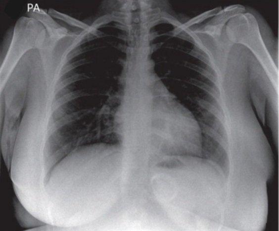
It's not a difficult case,but considered Must-know esp for professionals in emergency ward!CXR of a young girl presented with vomiting from 10 hours ago or so,also swallowing difficulty for almost anything.Other than an obesity surgery 3 years ago,no other diseases.
Dx & plan? Hint👉 This case has a clear-cut answer. I mean if you spot the abnormality correctly ,you already know how to manage this patient.
May 4, 2022 8 tweets 3 min read
🔴This tweet is not medical-related ! It's not about religion! If you're not interested in Quantum mechanics skip this,otherwise it may challenge your intuition at face value!

🔴So,is there really a distinction between the past,present,and future or could it all be in the mind? Image 🔴Part 1
Imagine traveling from City A to City B, you don't say that City B doesn’t exist because you aren't there yet!City B is a location in spacetime whether you're there or not.The same idea can be applied to time!So the future isn't going to happened, it is already there!
May 4, 2022 6 tweets 2 min read
These frontal & lateral views belong to a patient with a history of lung cancer which presented with a newly developed dyspnea. Considering the patient’s history and CXR findings,what is the most likely cause of the elevation of the right hemidiaphragm?
#chestrad #radiology Image Hint👉 I wanted to discuss about this subject and I incidentally encountered this case! Honestly speaking it's not complicated case ,but the question is worthy of noticing!
So do you think atelectasis of the R.U lobe is the main culprit?The answer based on many references is Not!