1/9 🧵 Age is the largest risk factor for Alzheimer’s disease. Our new study uses deep learning to try and understand metabolic and structural brain aging in this context. #endalz #AI @NaipMayo @MayoClinicNeuro 


2/9 We used both structural MRI (MAE 3.5 yrs) and FDG-PET (MAE 3.1 yrs) for deep learning based brain age prediction. FDG-PET is slightly better and both results had out-of-sample replication in ADNI. 

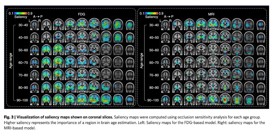
3/9 Saliency mapping showed clear difference in regions used for metabolic vs. structural brain age predictions. 

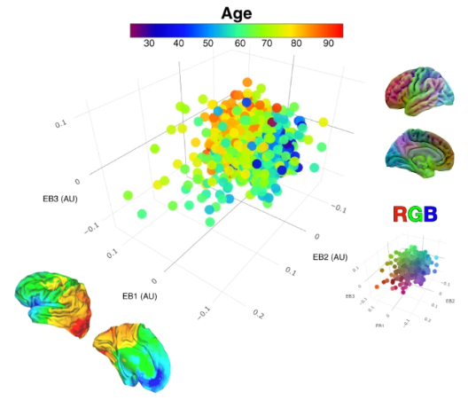
4/9 The posterior-to-anterior gradient in the saliency maps for FDG-based age prediction replicates the age relationship observed in our recent eigenbrain decomposition of FDG-PET. nature.com/articles/s4146… 

5/9 Corrected brain age gap (predicted age – actual age, corrected for age) is higher in AD and related disorders. The remaining age correlation with younger onset cases, is c/w more extreme disease phenotype as in other modalities (e.g., tau-PET levels in younger onset AD). 

6/9 FDG-based brain age gap was correlated with MRI-based brain age gap, but each also contained unique information. 

7/9 This interpretation lines up with the strong relationship between brain age gap and tau-PET in AD. 

8/9 Check out the paper to see how it relates to longitudinal cognitive decline, and how the brain age gap in AD looks more like accelerated aging than DLB or FTD. This converges with what we see in fMRI for AD and the DMN: ncbi.nlm.nih.gov/pmc/articles/P…
9/9 Tremendous effort lead by twitterless Jeyeon Lee! Much more to come from him and @NaipMayo !
Paper:
rdcu.be/cNbFX
Preprint: researchsquare.com/article/rs-804…
Paper:
rdcu.be/cNbFX
Preprint: researchsquare.com/article/rs-804…
• • •
Missing some Tweet in this thread? You can try to
force a refresh


