📕Month Review on Fabry Disease (FD) via @RCMjournal
🟡Mechanisms Beyond Storage & Forthcoming Therapies
🟡Cardiac Imaging
🟡Echocardiography #echofirst
🟡Cardiac Magnetic Resonance #WhyCMR
📂OPEN LINKS⬇️ & Thread🧵(1/13)
@mauripieroni72 @torresviera @SVCardio @DeBakeyCVedu
🟡Mechanisms Beyond Storage & Forthcoming Therapies
🟡Cardiac Imaging
🟡Echocardiography #echofirst
🟡Cardiac Magnetic Resonance #WhyCMR
📂OPEN LINKS⬇️ & Thread🧵(1/13)
@mauripieroni72 @torresviera @SVCardio @DeBakeyCVedu

✅FD X-Linked inherited Lysosomal Storage disorder
✅Mutations (>900) alfa-GAL gene (GLA)
✅🚫or⬇️ alfa-GAL A enzyme activity
✅Incidence 1/40,000-1/117,000
✅Newborn Screening 🇮🇹🇹🇼 1/8,800
✅FD storage GB3
✅Mutations (>900) alfa-GAL gene (GLA)
✅🚫or⬇️ alfa-GAL A enzyme activity
✅Incidence 1/40,000-1/117,000
✅Newborn Screening 🇮🇹🇹🇼 1/8,800
✅FD storage GB3

✅Intracellular glycosphingolipids organize➡️concentric lamellar bodies (🦓bodies)
✅lysoGB3➡️Pathogenic factor
✅Ion Channel Dysfunction
✅⬆️conduction velocity (atrial 🫀ventricular🫀)➡️short PR in absence of an accessory pathway

✅lysoGB3➡️Pathogenic factor
✅Ion Channel Dysfunction
✅⬆️conduction velocity (atrial 🫀ventricular🫀)➡️short PR in absence of an accessory pathway


✅Initial Phase➡️Start Childhood, 🫀storage without inflammation nor overt LVH
✅Second Phase➡️low T1 & high T2, 🧪Troponin & NT-proBNP
✅Third Phase➡️Marked LVH & Fibrosis
✅Amiodarone➡️worsen lysosomal pH⬇️effect of ERT (limited to selected cases with close monitoring)
✅Second Phase➡️low T1 & high T2, 🧪Troponin & NT-proBNP
✅Third Phase➡️Marked LVH & Fibrosis
✅Amiodarone➡️worsen lysosomal pH⬇️effect of ERT (limited to selected cases with close monitoring)

✅🫀most frequently affected organ >50% FD
✅Specific Tx➡️ERT💉(2001) & Chaperone💊(2016)
✅Data from FOS diagnosis➡️13.7y after onset of symptoms🧔♂️& 16.3y👩🦰
✅Specific Tx➡️ERT💉(2001) & Chaperone💊(2016)
✅Data from FOS diagnosis➡️13.7y after onset of symptoms🧔♂️& 16.3y👩🦰

✅Hallmark➡️LVH typically➡️concentric & without LVOTO
✅Binary Sign➡️Hyperechogenic region endocardial surface: black & white interface (not specific)
✅Papillary Hypertrophy
✅RVH without RV dysfunction

✅Binary Sign➡️Hyperechogenic region endocardial surface: black & white interface (not specific)
✅Papillary Hypertrophy
✅RVH without RV dysfunction


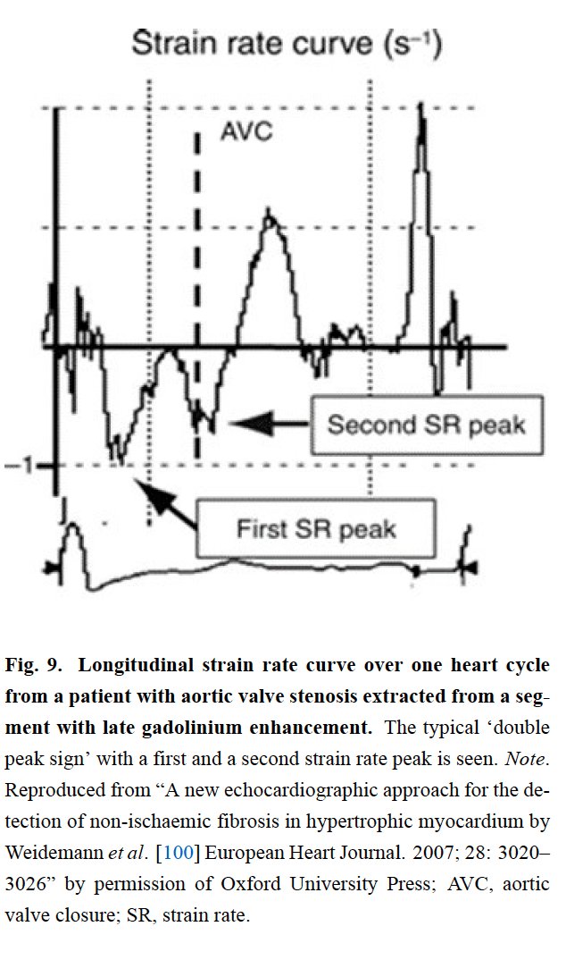
✅Reduction LS basal infero-lateral Wall (BILW)
✅BILW mos affected segment
✅Subepicardial layers most affected and earliest to occur
✅BILW mos affected segment
✅Subepicardial layers most affected and earliest to occur

✅Infiltration or Storage of Sphingolipids (T1)
✅Edema or Inflammation (T2)
✅Fibrosis (LGE)
✅Differential Dx FD with other causes LVH


✅Edema or Inflammation (T2)
✅Fibrosis (LGE)
✅Differential Dx FD with other causes LVH


✅Microvascular/Pre-accumulation Stage
✅Accumulation Stage
✅Inflammation/Hypertrophy Stage
✅Fibrosis/Impairment Stage
✅Severity LHV♂️("true LVH")>♀️("storage LVH")
✅♀️Preserved GLS until LVH♂️⬇️GLS & T1 before LVH
✅Inflammation/Fibrosis can precede LVH♀️rare♂️


✅Accumulation Stage
✅Inflammation/Hypertrophy Stage
✅Fibrosis/Impairment Stage
✅Severity LHV♂️("true LVH")>♀️("storage LVH")
✅♀️Preserved GLS until LVH♂️⬇️GLS & T1 before LVH
✅Inflammation/Fibrosis can precede LVH♀️rare♂️


✅#Echofirst FAST/non-invasive/low-cost/widely available/easy applicable & reproducible
✅Monitor disease/Estimate severity/Assess progression/Complications/Monitor treatment
✅LVH >12mm➡️Criterion start ERT
✅Monitor disease/Estimate severity/Assess progression/Complications/Monitor treatment
✅LVH >12mm➡️Criterion start ERT

✅Low T1➡️🫀glycosphingolipid storage
✅Low T1➡️early morphological alterations
✅"pre-storage" Phenotype➡️🫀trabeculations/early impairment in stress myocardial blood flow/subtle ECG abnormalities


✅Low T1➡️early morphological alterations
✅"pre-storage" Phenotype➡️🫀trabeculations/early impairment in stress myocardial blood flow/subtle ECG abnormalities


✅LGE➡️Basal infero-lateral Wall (75%)/mid-wall pattern
✅LGE➡️Marker advanced cardiac damage & poor response to ERT

✅LGE➡️Marker advanced cardiac damage & poor response to ERT


• • •
Missing some Tweet in this thread? You can try to
force a refresh


