
Thread alert!
In this thread, let us learn a little more about the imaging approach to post-cholecystectomy complications.
#RGphx
@cookyscan1
#Thread
#Twittorial
1/12
In this thread, let us learn a little more about the imaging approach to post-cholecystectomy complications.
#RGphx
@cookyscan1
#Thread
#Twittorial
1/12
https://twitter.com/RadioGraphics/status/1569380328908767233

The latest @RadioGraphics article by @EKKpdx and her team at @OHSURadiology focuses on multimodality imaging of post-cholecystectomy complications and their management.
Link: bit.ly/3RACwQK
#RGphx @CookyScan #tweetorial
2/12
Link: bit.ly/3RACwQK
#RGphx @CookyScan #tweetorial
2/12

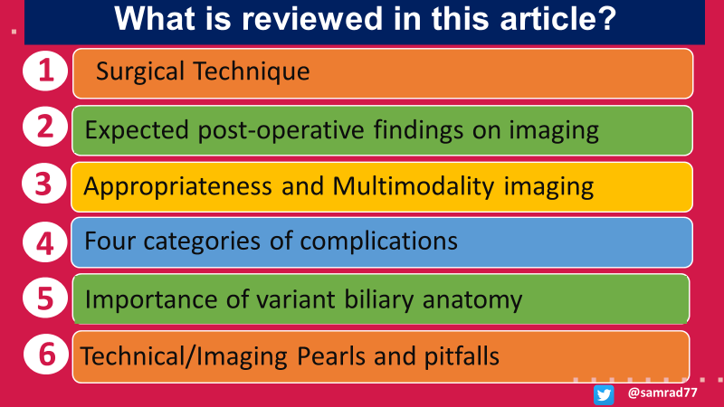
Cholecystectomy is one of the most performed surgeries. Radiologists and surgeons will, therefore, inevitably encounter several post-cholecystectomy complications. This article reviews role of imaging in diagnosing, preventing, and managing surgical complications.
#RGphx
3/12
#RGphx
3/12
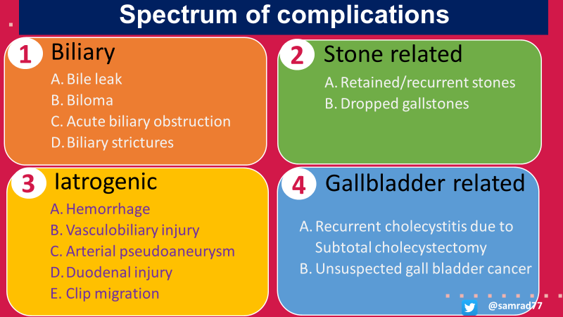
The authors conceptually organize post-cholecystectomy complications into four groups: biliary, stone-related, iatrogenic, and gallbladder-related.
#RGphx
4/12
#RGphx
4/12
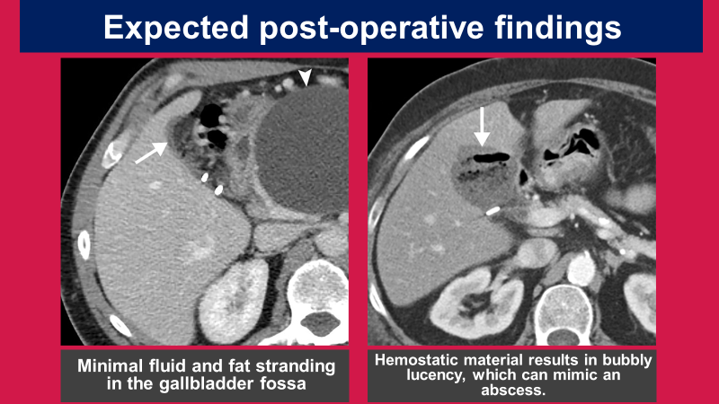
Expected post-operative findings on imaging include hyperechoic fat in the gall bladder fossa on ultrasound and a small amount of non-organized fluid-edema on USG/CT. Bubbly gas lucency of hemostatic material on CT shouldn’t be mistaken for a gas-forming infection.
#RGPhx
5/12
#RGPhx
5/12
Variant biliary anatomy is a risk factor for potential biliary injury. The two most important variants of surgical importance are the right posterior hepatic duct draining into the common hepatic or cystic duct and a subvesical bile duct (duct of Luschka).
#RGPhx
6/12
#RGPhx
6/12

Cystic variants (approximately 20%) of surgical importance are a low insertion of the cystic duct, a long parallel course of the cystic duct to the common hepatic duct, and a short cystic duct.
#RGPhx
7/12
#RGPhx
7/12

Bile leak results in an asymmetrical abdominal fluid or loculated collection with enhancing walls, which is often seen around the liver with/without communication with the gall bladder fossa.
#RGPhx
8/12
#RGPhx
8/12

99mTc HIDA scintigraphy with SPECT/CT, MRCP with gadoxetate disodium (Eovist/Primovist)–enhanced MRI, PTC or ERCP can confirm the bile leak and can show the site of the leak.
#RGPhx
9/12
#RGPhx
9/12

Dropped gallstones are essential to recognize as they can mimic mass or malignancy on imaging. Any unexplained abscess or mass in the hepatorenal fossa after cholecystectomy is highly suspicious for dropped stone-related complications.
#RGPhx
10/12
#RGPhx
10/12

Vascular injuries can result in hemorrhage in the gall bladder fossa or pseudoaneurysm. Multiphasic CT can diagnose hemorrhage and shows an active bleed or pseudoaneurysm.
#RGPhx
11/12
#RGPhx
11/12

In summary, the salient teaching points of this article are...
12/12
12/12

Thanks for coming along on the journey! Check out the excellent full-length @RadioGraphics article by @EKKpdx and her team at @OHSURadiology with plenty of practical tips and pearls to diagnose myriad post-cholecystectomy complications.
Link: bit.ly/3RACwQK
#RGphx
Link: bit.ly/3RACwQK
#RGphx

• • •
Missing some Tweet in this thread? You can try to
force a refresh


