Lea Alhilali, MD Profile picture
Oct 18, 2022 5 tweets 3 min read Read on X
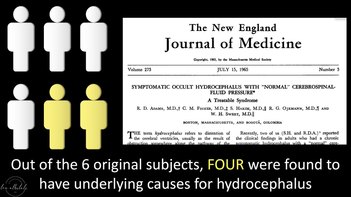
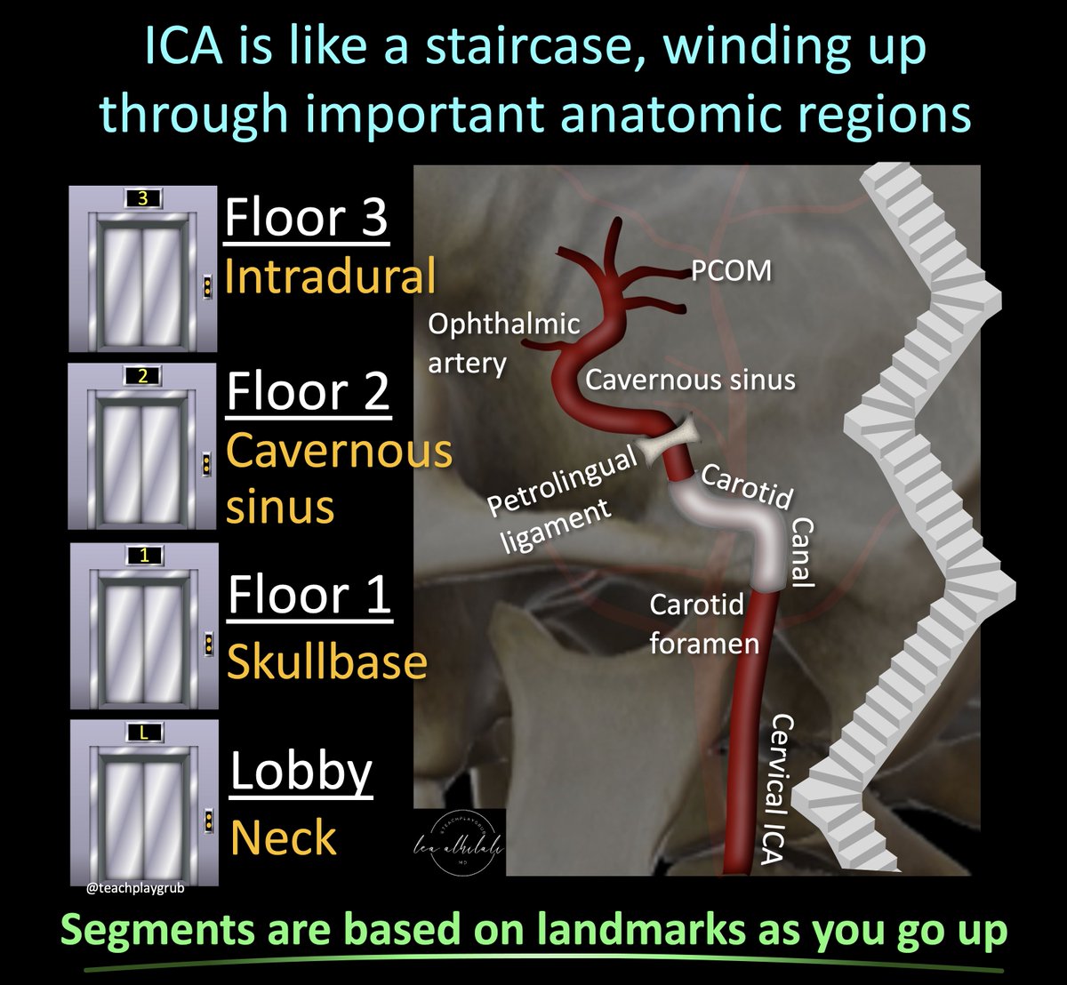
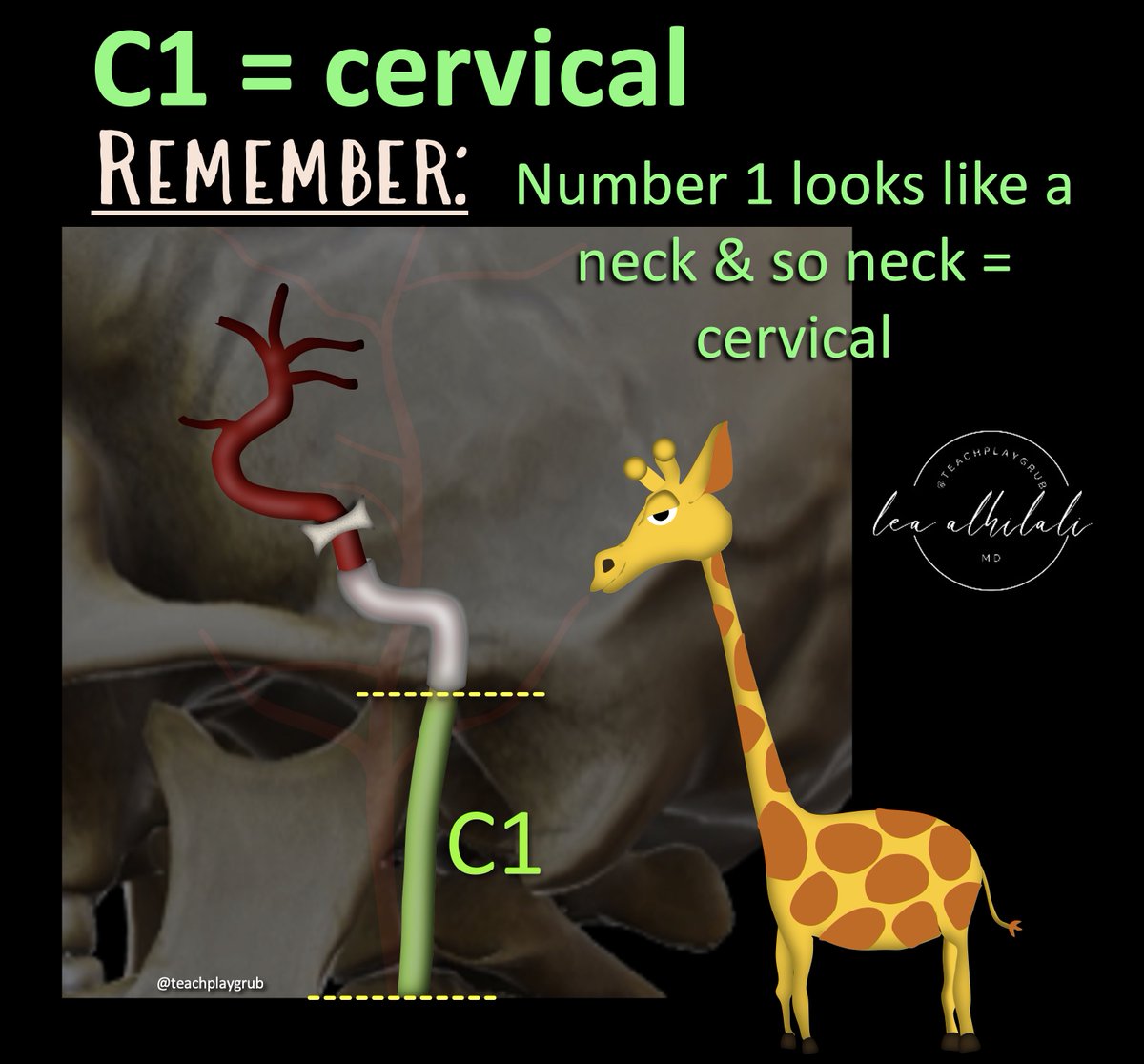
1/You don't need fancy programs to make great #Illustrations!

#Powerpoint is actually an extremely powerful illustrating tool--if you know the secret #hacks!

Here is a 🧵of videos explaining my best tips & tricks so you can make your own great figures, all just w/powerpoint! Image
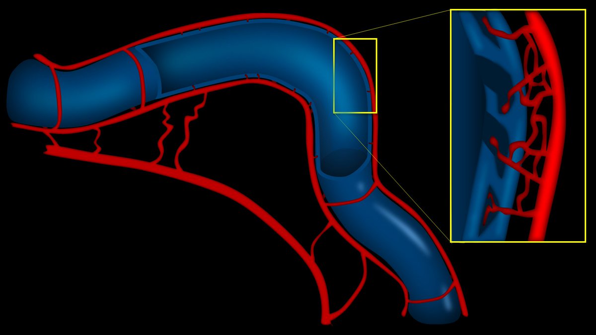
2/First, how to make a great 3D vessel in just 3 steps!

The video below shows you that all you need is a gradient, an edge shadow, and an inner glow to make an amorphous curve into a great figure of a vessel:

Image
3/Next, how to get a metallic sheen.

To make metal look real & 3D, you need that bright line of metallic reflection.

The video below shows you how to make a bland colored object reflect light like it's pure gold, just using simple gradients:

Image
4/And of course, one of PPT's most powerful drawing functions isn't even known by most people--the inner shadow function!

It makes figures of humans/bodies look more 3D.

The video below shows how to add 3D shadows right where you need it automatically

Image
5/I am working on making more videos on my powerpoint drawing tips, tricks, & hacks. So keep following for more!

Remember, you don't need expensive programs to make amazing figures for your presentation. You just need powerpoint, some quick tricks, and your own imagination!

• • •

Missing some Tweet in this thread? You can try to force a refresh
 

Keep Current with Lea Alhilali, MD

Lea Alhilali, MD Profile picture

Stay in touch and get notified when new unrolls are available from this author!

Read all threads

This Thread may be Removed Anytime!

PDF

Twitter may remove this content at anytime! Save it as PDF for later use!

Try unrolling a thread yourself!

how to unroll video
  1. Follow @ThreadReaderApp to mention us!

  2. From a Twitter thread mention us with a keyword "unroll"
@threadreaderapp unroll

Practice here first or read more on our help page!

More from @teachplaygrub

Jul 2
1/The medulla is anything but DULL!

Does seeing an infarct in the medulla cause your heart to skip a beat?

Does medullary anatomy send you into respiratory arrest?

Never fear, here is a thread on the major medullary syndromes! Image
2/The medulla is like a toll road.

Everything going down into the cord must pass through the medulla & everything from the cord going back up to the brain must too.

That’s a lot of tracts for a very small territory. Luckily you don’t need to know every tract Image
3/Medulla has 4 main vascular territories, spread out like a fan: anteromedial, anterolateral, lateral, and posterior.

You don’t need to remember their names, just the territory they cover—and I’ll show you how Image
Read 18 tweets
Jun 30
1/Time is brain! But what time is it?

If you don’t know the time of stroke onset, are you able to deduce it from imaging?

Here’s a thread to help you date a stroke on MRI! Image
2/Strokes evolve, or grow old, the same way people evolve or grow old.

The appearance of stroke on imaging mirrors the life stages of a person—you just have to change days for a stroke into years for a person

So 15 day old stroke has features of a 15 year old person, etc. Image
3/Initially (less than 4-6 hrs), the only finding is restriction (brightness) on diffusion imaging (DWI).

You can remember this bc in the first few months, a baby does nothing but be swaddled or restricted. So early/newly born stroke is like a baby, only restricted Image
Read 10 tweets
Jun 27
1/”I LOVE spinal cord syndromes!” is a phrase that has NEVER, EVER been said by anyone.

Do you become paralyzed when you see cord signal abnormality?

Never fear—here is a thread on all the incomplete spinal cord syndromes to get you moving again! Image
2/Spinal cord anatomy can be complex. On imaging, we can see the ant & post nerve roots. We can also see the gray & white matter. Hidden w/in the white matter, however, are numerous efferent & afferent tracts—enough to make your head spin. Image
3/Lucky for you, for the incomplete cord syndromes, all you need to know is gray matter & 3 main tracts. Anterolaterally, spinothalamic tract (pain & temp). Posteriorly, dorsal columns (vibration, proprioception, & light touch), & next to it, corticospinal tracts—providing motor Image
Read 20 tweets
Jun 23
1/Do you get a Broca’s aphasia trying remember the location of Broca's area?

Does trying to remember inferior frontal gyrus anatomy leave you speechless?

Don't be at a loss for words when it comes to Broca's area

Here’s a 🧵to help you remember the anatomy of this key region! Image
2/Anatomy of the inferior frontal gyrus (IFG) is best seen on the sagittal images, where it looks like the McDonald’s arches.

So, to find this area on MR, I open the sagittal images & scroll until I see the arches. When it comes to this method of finding the IFG, i’m lovin it. Image
3/Inferior frontal gyrus also looks like a sideways 3, if you prefer. This 3 is helpful bc the inferior frontal gyrus has 3 parts—called pars Image
Read 13 tweets
Jun 19
1/Feeling intoxicated trying to remember all the findings in alcohol use disorder?!

Here’s something to put you in high spirits!

This month’s @Radiographics has the important neuroimaging findings alcohol use disorder!


@cookyscan1 @RadG_editor #RGphx pubs.rsna.org/doi/10.1148/rg…Image
2/There’s an easy rhyme to help you remember the important neuroimaging findings of alcohol use disorder

“Basal ganglia is white...”

Get intrinsic T1 shortening in the BG that makes it look white as a ghost! Image
3/Next “...Cortex is bright”

Acute hyperammonemic encephalopathy cause cortical restricted diffusion, especially the insula, so that it looks as bright as a light bulb! Image
Read 8 tweets
Jun 9
1/Need help reading spine imaging? I’ve got your back!

It’s as easy as ABC!

A thread about an easy mnemonic you can use on every single spine study you see to increase your speed & make sure you never miss a thing! Image
2/A is for alignment

Look for:
(1) Unstable injuries

(2) Malalignment that causes early degenerative change. Abnormal motion causes spinal elements to abnormally move against each other, like grinding teeth wears down teeth—this wears down the spine Image
3/B is for bones.

On CT, the most important thing to look for w/bones is fractures. You may see focal bony lesions, but you may not

On MR, it is the opposite—you can see marrow lesions easily but you may or may not see edema associated w/fractures if the fracture is subtle Image
Read 11 tweets

Did Thread Reader help you today?

Support us! We are indie developers!


This site is made by just two indie developers on a laptop doing marketing, support and development! Read more about the story.

Become a Premium Member ($3/month or $30/year) and get exclusive features!

Become Premium

Don't want to be a Premium member but still want to support us?

Make a small donation by buying us coffee ($5) or help with server cost ($10)

Donate via Paypal

Or Donate anonymously using crypto!

Ethereum

0xfe58350B80634f60Fa6Dc149a72b4DFbc17D341E copy

Bitcoin

3ATGMxNzCUFzxpMCHL5sWSt4DVtS8UqXpi copy

Thank you for your support!

Follow Us!

:(