Kristen Scheitler, MD Profile picture
Nov 16, 2022 9 tweets 4 min read Read on X
1/ Have you ever had a patient with low back pain & degenerative findings on lumbar MR imaging? 🔎👀

Demystifying lumbar stenosis is one of my favorite things to teach medical students & junior residents!

A thread 🧵
2/ Let’s review the normal anatomy of the L3-4 spinal segment.

🟡 Mid-sagittal: we see the central canal w/ the thecal sac, containing nerve roots distal to the conus.

🟠 Parasagittal: the lumbar nerve roots exit below their respective pedicles via the neural foramina.
3/ Normal anatomy cont’d:

🔵 Posterior view: we see the relationship between “exiting” nerve roots & their respective pedicles, as well as “traversing” nerve roots.

⚪️ Axial (simplified): shows the relationship between the disc, thecal sac, root, and joint.
4/ “Lumbar stenosis” refers to anything that decreases the area of:

▫️central canal (thecal sac)
▫️lateral recess
▫️neural foramen

Correspondingly, we classify pathologies anatomically as causing “central,” “lateral recess,” or “foraminal” stenosis (color-coded below).
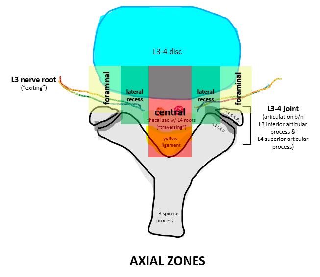
5/ Central stenosis 🟰 compression of the thecal sac by any central pathology (central disc herniation, yellow ligament hypertrophy, epidural lipomatosis, &c).

Primary symptoms:

🔻neurogenic pseudoclaudication
🔻traversing (L4) nerve root radicular pain/numbness/or weakness
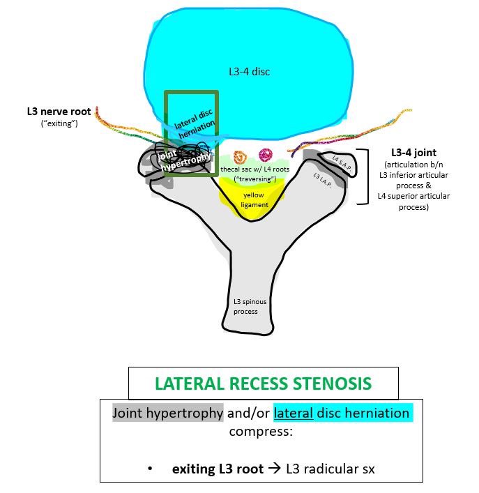
6/ Lateral recess stenosis 🟰 compression in the area bound by the pedicle, vertebral body, & superior articular process…basically the area containing the first part of the exiting root (L3).

Most common symptoms:

🔻exiting (L3) nerve root radicular pain/numbness/or weakness
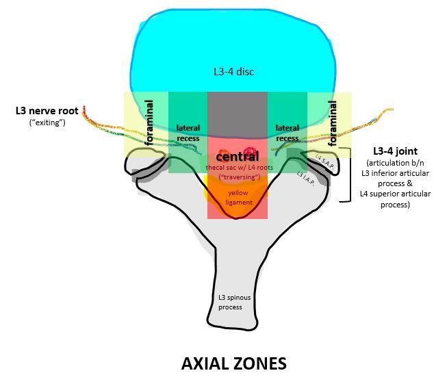
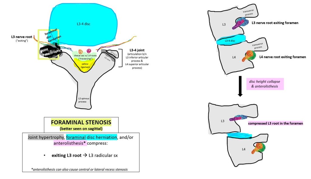
7/ Foraminal stenosis = compression of the exiting nerve root in the foramen, distal to the lateral recess. Common causes include lateral disc herniation, joint hypertrophy, spondylolisthesis, &c.

Main symptoms:

🔻exiting (L3) nerve root radicular pain/numbness/or weakness
8/ SUMMARY: Lumbar stenosis has many causes & is classified anatomically. Each classification has corresponding implications for symptom localization & treatment!

Disclaimer: content was simplified for purposes of conceptualization.

Any comments or extra info welcomed :)
🙏 You can find my thread on cauda equina syndrome here:

• • •

Missing some Tweet in this thread? You can try to force a refresh
 

Keep Current with Kristen Scheitler, MD

Kristen Scheitler, MD Profile picture

Stay in touch and get notified when new unrolls are available from this author!

Read all threads

This Thread may be Removed Anytime!

PDF

Twitter may remove this content at anytime! Save it as PDF for later use!

Try unrolling a thread yourself!

how to unroll video
  1. Follow @ThreadReaderApp to mention us!

  2. From a Twitter thread mention us with a keyword "unroll"
@threadreaderapp unroll

Practice here first or read more on our help page!

More from @k_scheitler

Apr 25, 2023
Need help remembering how to compute a Glasgow Coma Scale (GCS) score?
 
A thread:

How to quickly and reliably compute GCS without having to look it up every time 😎👇🧵🧠

/1
The Glasgow Coma Scale (GCS) was first published in the 1970s and was intended to describe a patient's level of consciousness following traumatic brain injury.

/2 Image
GCS has 3 components:
 
👀 Eye-opening (4 points)
👄 Verbal (5 pts)
💪 Motor (6 pts)
 
Don't memorize this!

When I was a student, someone taught me to remember this simply as:
 
EYES (4 letters = 4 total pts)
WORDS (5 letters = 5 total pts)
MOVING (6 letters = 6 total pts)

/3
Read 12 tweets
Mar 24, 2023
General principles of cranial stabilization for neurosurgical procedures – a thread 🧵

/1
Intraoperative head stabilization is often performed by pinning. Careful & thoughtful consideration is required to
 
-avoid complications
-optimize access to surgical target & minimize steric hindrances
-support devices for stereotactic neuronavigation or retraction

/2
First, review neurosurgical hx & imaging:
 
-Any prior craniotomy/hardware/implants (e.g., shunt, DBS leads, etc)
-Relevant anatomy (e.g., frontal sinus, mastoid aeration, skull thinning from chronic hydrocephalus)
-Need for future bypass/donor scalp vessel (e.g, STA or OA)?

/3
Read 8 tweets
Mar 16, 2023
External ventricular drains (EVDs) are one of the most common procedures in neurosurgery… And having to troubleshoot said EVDs that stop working (usually in the middle of the night) is ALSO common, though not always intuitive 📟🫠

How to troubleshoot an EVD - a thread 🧵 /1
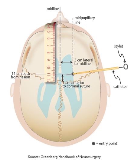
First, let's review how EVDs work. An EVD is a temporary catheter placed in the ventricle at the foramen of Monro that can (1) measure ICP & (2) control ICP by draining CSF.

/2
The drainage system is leveled at the ear, and the collection chamber is raised to different heights to control CSF flow and, thus, drainage.

At a given height, CSF will drain whenever intraventricular pressure exceeds that set by the height of the collection system.

/3
Read 9 tweets
Aug 22, 2022
You're paged emergently about an incoming patient with a suspected brain injury.

What are the first things you need to do?

A review of the emergent evaluation of a patient with traumatic epidural or subdural hematoma
🧵🧠👇
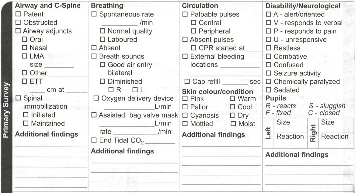
1. Advanced Trauma Life Support (ATLS) primary survey (airway, breathing, circulation, etc.).

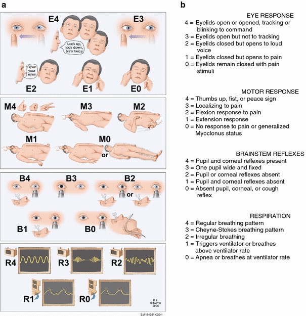
2. Rapid neurologic assessment with a validated coma scale (either GCS or FOUR score). Prior to exam, confirm paralytic reversal with train of fours and pause sedation. Image
Image
Image
3. Emergent non-contrast CT head, with close attention to any accompanying skull base fractures and parenchymal signs of increased ICP (i.e., sulcal effacement, effacement of basal cisterns, herniation, etc). Image
Image
Image
Read 9 tweets
May 13, 2022
One of the most common consults we see in neurosurgery is the 'cauda equina syndrome (CES) rule-out.' CES can be diagnostically challenging & panic-inducing due to its highly variable presentation & grave consequences if missed.

How to evaluate suspected CES: a thread 🧵

(1/9)
(2/9) When cauda equina syndrome is suspected, investigate the following:

•History (always)
•Bladder function (PVR)
•Rectal exam (3 parts: perianal sensation, voluntary contraction, wink reflex)
•Neuro exam (sensorimotor, reflexes)
•Imaging (MRI)
 
Let’s dive in 👇
(3/9) We all learned:

bladder/bowel dysfunction + saddle anesthesia + severe leg/low back pain = slam-dunk diagnosis

...but the presentation is not always this clear.

Fortunately, cauda equina syndrome is RARE - annual incidence is approx. 5-10 cases per 1,000,000.
Read 9 tweets
May 11, 2022
Elie Wiesel gave the commencement speech at my college graduation - 'Memory and Ethics.' His words are as germane as ever. A thread 🧵

"One thing I can tell you: You see that road there? Don’t go there. I have just come from there..." (1/7)

(2/7) "...I belong to a generation that tells you that. The 20th century was one of the worst centuries in the history of humankind. Why? Because it was dominated by two fanaticisms. Political fanaticism. Racist fanaticism. That century caused more deaths than any time before."
(3/7) "...What do we know now? A new trend is hanging upon us, and the name is fanaticism. We must do whatever we can to, first of all, unmask. Second, to denounce. And, of course, to oppose fanaticism wherever it is."
Read 7 tweets

Did Thread Reader help you today?

Support us! We are indie developers!


This site is made by just two indie developers on a laptop doing marketing, support and development! Read more about the story.

Become a Premium Member ($3/month or $30/year) and get exclusive features!

Become Premium

Don't want to be a Premium member but still want to support us?

Make a small donation by buying us coffee ($5) or help with server cost ($10)

Donate via Paypal

Or Donate anonymously using crypto!

Ethereum

0xfe58350B80634f60Fa6Dc149a72b4DFbc17D341E copy

Bitcoin

3ATGMxNzCUFzxpMCHL5sWSt4DVtS8UqXpi copy

Thank you for your support!

Follow Us!

:(