Meningeal signs
“Brudziński neck sign, Brudziński symphyseal sign, Brudziński cheek sign, Brudzinski's reflex”
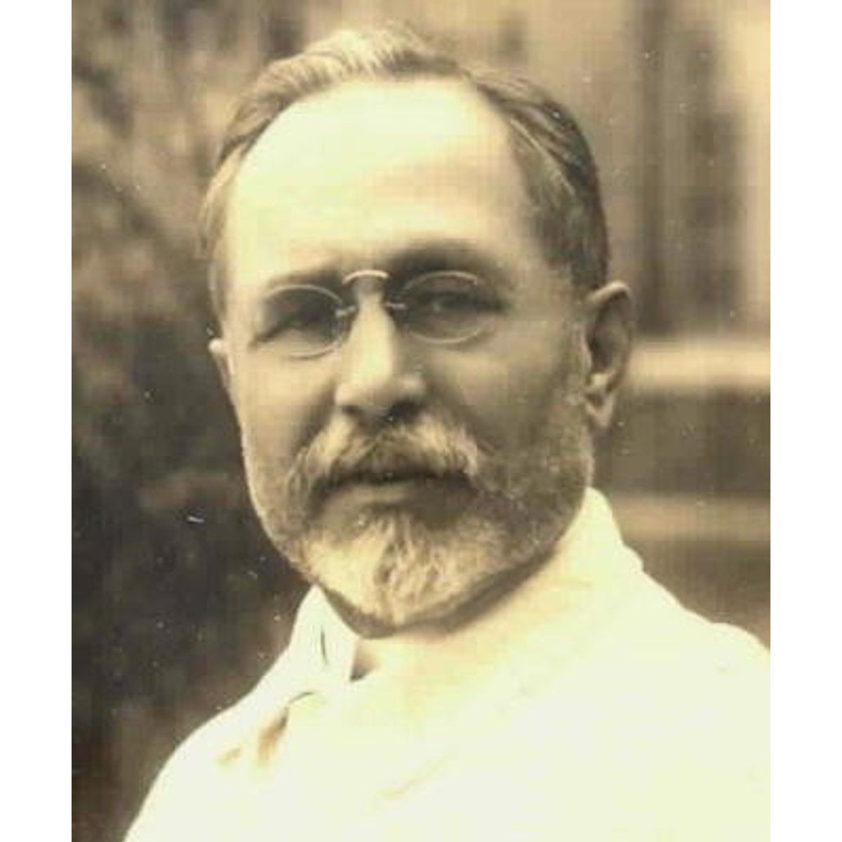
Polish pediatrician Józef Polikarp Brudziński (1874–1917)
#MedTwitter #neurotwitter #EndNeurophobia #tweetorials
1/
“Brudziński neck sign, Brudziński symphyseal sign, Brudziński cheek sign, Brudzinski's reflex”
Polish pediatrician Józef Polikarp Brudziński (1874–1917)
#MedTwitter #neurotwitter #EndNeurophobia #tweetorials
1/

Definitions
Meningitis
-inflammation of leptomeninges & underlying subarachnoid CSF
Meningismus
-morbid state characterized by meningitic syndrome (triad: headache, photophobia, nuchal rigidity)
Meningism
-synonymous of meningismus
-neck stiffness w/o meningeal inflammation
2/
Meningitis
-inflammation of leptomeninges & underlying subarachnoid CSF
Meningismus
-morbid state characterized by meningitic syndrome (triad: headache, photophobia, nuchal rigidity)
Meningism
-synonymous of meningismus
-neck stiffness w/o meningeal inflammation
2/
Mechanism
“maneuvers used to elicit meningeal signs produce tension on inflamed and hypersensitive spinal nerve roots, and the resulting signs are postures, protective muscle contractions, or other movements that minimize the stretch and distortion of the meninges and roots”
3/
“maneuvers used to elicit meningeal signs produce tension on inflamed and hypersensitive spinal nerve roots, and the resulting signs are postures, protective muscle contractions, or other movements that minimize the stretch and distortion of the meninges and roots”
3/
Diagnostic value
a. Kernig's sign (Thomas et al 2002)
SN 5% SP 95% PPV 27% NPV 72%
b. absence of fever, neck stiffness, and altered mental status (DeJong)
exclude meningitis (SN 99%)
4/
a. Kernig's sign (Thomas et al 2002)
SN 5% SP 95% PPV 27% NPV 72%
b. absence of fever, neck stiffness, and altered mental status (DeJong)
exclude meningitis (SN 99%)
4/
Maneuvers used to elicit meningeal signs
a. Nuchal (cervical) rigidity
b. Kernig's sign
c. Brudzinski's neck sign
d. Brudzinski's contralateral reflex sign
e. Tripod or Amoss's or Hoyne's sign
f. Others
5/
a. Nuchal (cervical) rigidity
b. Kernig's sign
c. Brudzinski's neck sign
d. Brudzinski's contralateral reflex sign
e. Tripod or Amoss's or Hoyne's sign
f. Others
5/
Nuchal rigidity
a. relax neck muscles by supine patient to edge of bed& allow head to hang outside bed
b. place your hand under patient's head & try to flex neck, touch chin to chest
c. resistance implies diffuse irritation of cervical nerve roots from meningeal
6/
a. relax neck muscles by supine patient to edge of bed& allow head to hang outside bed
b. place your hand under patient's head & try to flex neck, touch chin to chest
c. resistance implies diffuse irritation of cervical nerve roots from meningeal
6/
Nuchal rigidity
via: Endeavour For Child Health
7/
via: Endeavour For Child Health
7/
Kernig's sign
a. flex hip&knee on 1-side while patient is supine
b. now, extend knee w/ hip still flexed
c. +, if hamstring spasm results in pain in post thigh & difficulty w/ knee extension
d. severe inflammation of meninges, the opposite knee may also flex
8/
a. flex hip&knee on 1-side while patient is supine
b. now, extend knee w/ hip still flexed
c. +, if hamstring spasm results in pain in post thigh & difficulty w/ knee extension
d. severe inflammation of meninges, the opposite knee may also flex
8/
Kernig's sign
via: Neuron Bundle
9/
via: Neuron Bundle
9/
Kernig's sign
via: Dr. Akash Gangane
10/
via: Dr. Akash Gangane
10/
Kernig's sign
via: Dr Aishwarya Kelkar Medical Lectures
11/
via: Dr Aishwarya Kelkar Medical Lectures
11/
Brudzinski's neck sign
a.supine patient, place one of your hand behind his head & other on his/her chest
b. now, flex patient’s head with hand behind head, while your hand on chest restrains patient and prevents him/her from rising
c.+, if the patient flexes hips and knees.
12/
a.supine patient, place one of your hand behind his head & other on his/her chest
b. now, flex patient’s head with hand behind head, while your hand on chest restrains patient and prevents him/her from rising
c.+, if the patient flexes hips and knees.
12/
Brudzinski's neck sign
via: Dr. Akash Gangane
13/
via: Dr. Akash Gangane
13/
Brudzinski's neck sign
via: Dr. Akash Gangane
14/
via: Dr. Akash Gangane
14/
Brudzinski's neck sign
via: MBBS VPASS
15/
via: MBBS VPASS
15/
Brudzinski's contralateral reflex
a.2 parts: identical&reciprocal contralateral reflex
b.identical: contralateral leg bends w/ passive flexion of patient's hip&knee on 1-side
c.reciprocal: +, when contralateral leg that has flexed reflexly begins to extend (little kick)
16/
a.2 parts: identical&reciprocal contralateral reflex
b.identical: contralateral leg bends w/ passive flexion of patient's hip&knee on 1-side
c.reciprocal: +, when contralateral leg that has flexed reflexly begins to extend (little kick)
16/
Brudzinski's contralateral reflex sign
via: Endeavour For Child Health
17/
via: Endeavour For Child Health
17/
Tripod, Amoss, Hoyne sign
Patient is asked to sit up in bed. Health, sits up w/o supporting himself
Meningeal irritation: patient tries to sit up by supporting himself w/ his hands placed far behind him in bed, to take the weight off the spine and prevent its flexion.
18/
Patient is asked to sit up in bed. Health, sits up w/o supporting himself
Meningeal irritation: patient tries to sit up by supporting himself w/ his hands placed far behind him in bed, to take the weight off the spine and prevent its flexion.
18/

Bikele sign
Positive, in meningeal inflammation and in brachial plexitis
19/
Positive, in meningeal inflammation and in brachial plexitis
19/

Josef Brudzinski discoveries
Polish pediatrician Josef Brudzinski who had described four different maneuvers for the diagnosis of meningitis in the early-20th century
Brudziński cheek sign
Brudziński symphyseal sign
Brudzinski's reflex
Brudzinski's sign
20/
Polish pediatrician Josef Brudzinski who had described four different maneuvers for the diagnosis of meningitis in the early-20th century
Brudziński cheek sign
Brudziński symphyseal sign
Brudzinski's reflex
Brudzinski's sign
20/

Guilland's sign
- pinching the skin over the quadriceps femoris muscle or squeezing the muscle on one side cause flexion of contralateral hip and knee
Edelmann great toe phenomenon
- flexion of the hip with the knee extended cause extension of the great toe
21/
- pinching the skin over the quadriceps femoris muscle or squeezing the muscle on one side cause flexion of contralateral hip and knee
Edelmann great toe phenomenon
- flexion of the hip with the knee extended cause extension of the great toe
21/
Binda sign
- passively alternating rapidly turning the head, the opposite shoulder is raised
Plantar neck
- Babinski when flexing the head
Flatau sign
- midriasis by Brudzinski’s sign
Parrot sign
- midriasis by skin pinch
22/
- passively alternating rapidly turning the head, the opposite shoulder is raised
Plantar neck
- Babinski when flexing the head
Flatau sign
- midriasis by Brudzinski’s sign
Parrot sign
- midriasis by skin pinch
22/
Skeer sign
- peripupillary rim (TB meningitis)
Magnus sign
- turning the head to the side, the patient extends the arm rostral and flexes caudal
Lewinson sign
- when asking the patient to touch his chest with his chin, he opens his mouth in an attempt to do so
23/
- peripupillary rim (TB meningitis)
Magnus sign
- turning the head to the side, the patient extends the arm rostral and flexes caudal
Lewinson sign
- when asking the patient to touch his chest with his chin, he opens his mouth in an attempt to do so
23/
Lafora sign
- hypersensitivity when gently stimulating the nasal mucosa
Signorelli sign
- hyperesthesia when performing the Foix maneuver
Arnos sign
- By asking the patient to get up from the supine position with his arms crossed, he cannot execute it
24/
- hypersensitivity when gently stimulating the nasal mucosa
Signorelli sign
- hyperesthesia when performing the Foix maneuver
Arnos sign
- By asking the patient to get up from the supine position with his arms crossed, he cannot execute it
24/
Trousseau sign (meningitic line)
- vasomotor phenomenon, imprint left by a finger or blunt object as it passes over the skin surface
Jolt sign
- headache accentuation, when patient is asked to turn his head horizontally 2-3 rotations/sec
25/
- vasomotor phenomenon, imprint left by a finger or blunt object as it passes over the skin surface
Jolt sign
- headache accentuation, when patient is asked to turn his head horizontally 2-3 rotations/sec
25/
Shotgun trigger
- classic trunk flexion position with neck hyperextension
Jacoud sign
- bradycardia by Brudzinski’s sign (TB meningitis)
Lesague sign
- When lifting a child by armpits, he remains in the air with his legs bent
26/
- classic trunk flexion position with neck hyperextension
Jacoud sign
- bradycardia by Brudzinski’s sign (TB meningitis)
Lesague sign
- When lifting a child by armpits, he remains in the air with his legs bent
26/
NeuroTeach - Content
The blog contains all the threads and videos.
neuronland.blogspot.com/2022/11/neurot…
Have a great day!
27/
The blog contains all the threads and videos.
neuronland.blogspot.com/2022/11/neurot…
Have a great day!
27/
• • •
Missing some Tweet in this thread? You can try to
force a refresh