The proper understanding and use of melatonin may not only aid those with 💉 injuries and/or Long COVID; but also those who are exposed to low-level microwaves, EMFs, 60 Hz magnetic field, and ambient light at night.
So pretty much everyone.
A THREAD 🧵
So pretty much everyone.
A THREAD 🧵
https://twitter.com/drkohilathas/status/1628344715715268610
In the original thread we learnt that melatonin has potent antioxidant and anti-inflammatory effects.
Due to this, it has frequently been proposed for use to overcome the cytokine storm in virus-related infections, including that caused by SARS-CoV-2.
link.springer.com/article/10.100…
Due to this, it has frequently been proposed for use to overcome the cytokine storm in virus-related infections, including that caused by SARS-CoV-2.
link.springer.com/article/10.100…
Melatonin has been put forward as a potential treatment aid for COVID-19 and long COVID for a number of other reasons too.
One of them is by shifting cellular energy production by quelling HIF-1α.
One of them is by shifting cellular energy production by quelling HIF-1α.
Life is energy, and good health is a state of proper energy efficiency.
When unwell with an infection, immune cells need higher energy demands more immediately and thus switch from getting energy from mitochondrial oxidative phosphorylation to cytosolic glycolysis.
When unwell with an infection, immune cells need higher energy demands more immediately and thus switch from getting energy from mitochondrial oxidative phosphorylation to cytosolic glycolysis.

Glycolysis is an oxygen-independent and relatively inefficient way to generate energy compared to oxidative phosphorylation but found to be the dominant metabolic pathway in pro-inflammatory cells.
One of many regulators that help this switch is hypoxia-inducible factor-1α (HIF-1α).
link.springer.com/article/10.100…
link.springer.com/article/10.100…
Increased mortality is observed in patients with elevated HIF-1α and is associated with severe cytokine release.
sciencedirect.com/science/articl…
sciencedirect.com/science/articl…
Melatonin is able to quell HIF-1α and switch cellular energy production back to the healthier mitochondrial oxidative phosphorylation.
mdpi.com/1422-0067/22/2…
mdpi.com/1422-0067/22/2…
Adenosine triphosphate (ATP) is the source of energy for use and storage at the cellular level, and melatonin is well recognised for its ability to protect and enhance ATP production in mitochondria.
ncbi.nlm.nih.gov/pmc/articles/P…
ncbi.nlm.nih.gov/pmc/articles/P…
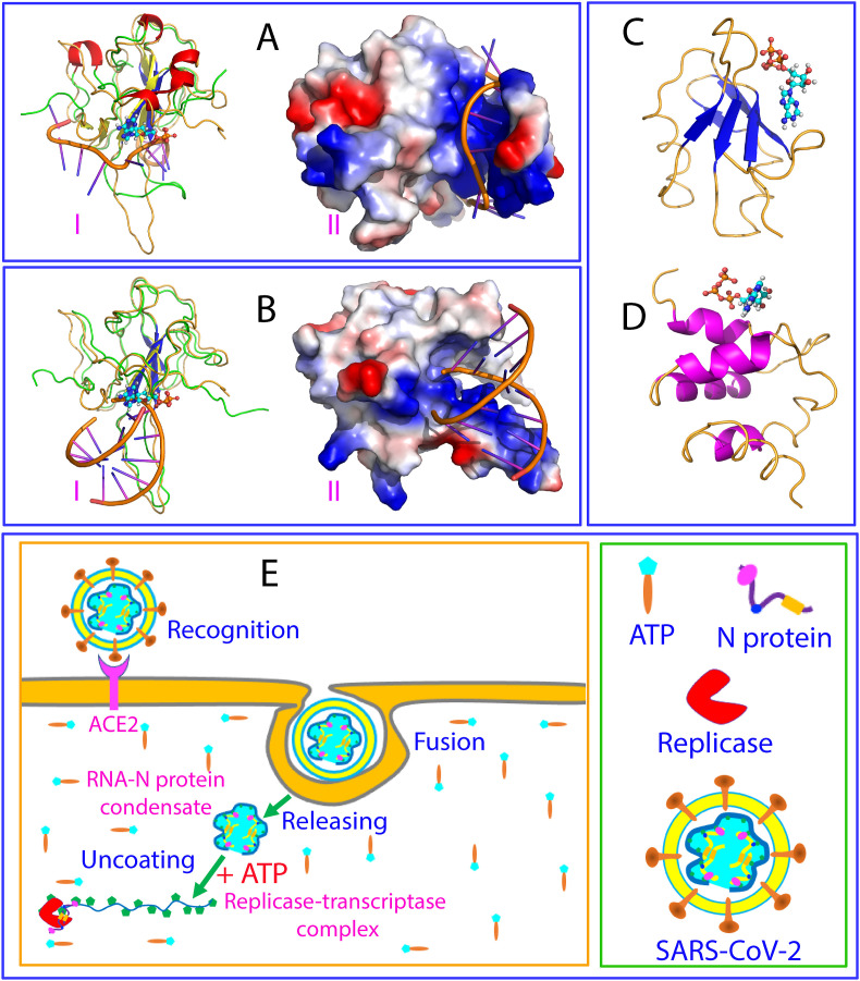
Other than making us move...and live, ATP is capable of completely dissolving viral factories in cells but only if it outnumbers viral proteins by many folds.
One study showed ATP was capable of completely dissolving viral condensates formed by SARS-CoV-2 N protein, but only at ratios of 1:500 (N-protein:ATP).
pubmed.ncbi.nlm.nih.gov/33477032/
pubmed.ncbi.nlm.nih.gov/33477032/
Higher ATP may thus reduce viral replication, which may explain why severe COVID-19 in children is rare.
Children have higher plasma levels of ATP that are negatively correlated with the frequency of regulatory T cells but positively correlated with the frequency of CD4+ T cells.
ncbi.nlm.nih.gov/pmc/articles/P…
ncbi.nlm.nih.gov/pmc/articles/P…
Energy efficiency is important, and hence why a low carbohydrate diet may help "boost" T cell function.
https://twitter.com/drkohilathas/status/1635251926639218688?s=20
Melatonin may upregulate ATP production and thus help with viral protection.
ncbi.nlm.nih.gov/pmc/articles/P…
ncbi.nlm.nih.gov/pmc/articles/P…
Melatonin can also change the pathological shape of mitochondria too.
Mitochondria infected by SARS-CoV-2 display swollen inner folds, this causes mitochondria to not work effectively preventing higher ATP production via oxidative phosphorylation in favour of glycolysis.
Mitochondria infected by SARS-CoV-2 display swollen inner folds, this causes mitochondria to not work effectively preventing higher ATP production via oxidative phosphorylation in favour of glycolysis.
The mitochondria of white blood cells in those recovered from COVID-19 have also shown dysfunction even at 11 months post-infection.
Damaged mitochondria continue to produce more ROS.
A vicious cycle is formed.
ncbi.nlm.nih.gov/pmc/articles/P…
Damaged mitochondria continue to produce more ROS.
A vicious cycle is formed.
ncbi.nlm.nih.gov/pmc/articles/P…
Melatonin and its metabolites are able to attenuate mitochondrial damage and are extremely effective at scavenging different types of ROS.
academic.oup.com/biomedgerontol…
academic.oup.com/biomedgerontol…
A lot of us are worried that many that have had the 💉 may have unknowingly integrated external external genetic material into the DNA is via a molecule called long interspersed element-1 (LINE-1 or L1) retrotransposons.
https://twitter.com/drkohilathas/status/1637795453214371840?s=20
Though coronavirus RNAs are not supposed to reverse-transcribe and integrate into host DNA, recent research found that, via LINE1, SARS-CoV-2 and other human coronaviruses could insert into the host genome.
ncbi.nlm.nih.gov/pmc/articles/P…
ncbi.nlm.nih.gov/pmc/articles/P…
Repression is a mechanism often used to decrease or inhibit the expression of a gene, and in healthy states, LINE-1 is repressed. 
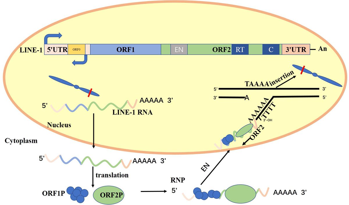
Removal of repression is called derepression, and LINE-1 derepression is linked to ageing, diabetes, cardiac abnormalities, mitochondrial dysfunction and cancer.
ncbi.nlm.nih.gov/pmc/articles/P…
ncbi.nlm.nih.gov/pmc/articles/P…
Melatonin may inhibit LINE1 expression via antioxidant-dependent and independent mechanisms.
ncbi.nlm.nih.gov/pmc/articles/P…
ncbi.nlm.nih.gov/pmc/articles/P…
Before we go further, we have to understand what liquid–liquid phase separation (LLPS) is.
Cells contain little factories called organelles that perform certain functions.
Some have an encapsulating membrane while others don't.
Cells contain little factories called organelles that perform certain functions.
Some have an encapsulating membrane while others don't.
A fundamental question in cell biology is
“how are these membraneless compartments organised to control such complex biochemical reactions in space and time.”
“how are these membraneless compartments organised to control such complex biochemical reactions in space and time.”

Evidence suggest that biomolecular condensates (or little pockets/bubbles within cells) are reversibly and dynamically assembled via LLPS.
LLPS is a process that spontaneously drives the separation of a homogeneous solution of constituents into two or more phases.
LLPS is a process that spontaneously drives the separation of a homogeneous solution of constituents into two or more phases.
Two types of multivalent interactions contribute to LLPS:
1. Intracellular protein-protein, protein–RNA, and RNA–RNA interactions.
2. Weak, transient, multivalent interactions between intrinsically disordered regions
1. Intracellular protein-protein, protein–RNA, and RNA–RNA interactions.
2. Weak, transient, multivalent interactions between intrinsically disordered regions
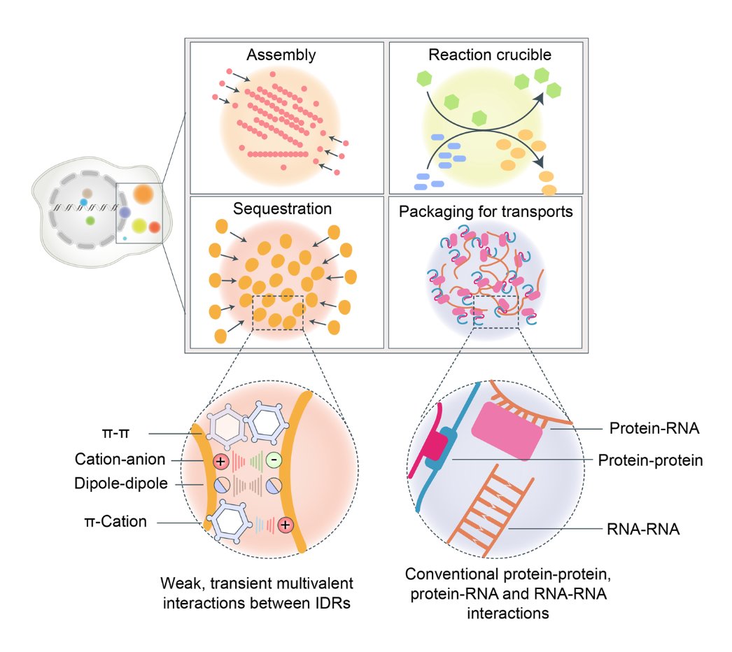
And so LPPS can be described as a product of the force of electrochemical gradients within cells, and these gradients are established by the multivalent interactions, which influence and are influenced by the spatial arrangement of molecules in the droplets.
Some physiological functions of LLPS include:
- Regulating transcription
- Being involved in genome organisation
- Being involved in the immune system
- Neuronal synaptic signalling
- Regulating transcription
- Being involved in genome organisation
- Being involved in the immune system
- Neuronal synaptic signalling
Dysregulated LLPS that occurs in aberrant cellular processes associated with carcinogenesis, including chromatin organisation, epigenetics, oncogenic transcription, aberrant signaling pathways, and telomere lengthening mediated by LLPS.
Pathological functions of LLPS include:
- Neurodegenerative disease - via pathological protein aggregates originate from an aberrant phase separation.
- Cancer - Beyond gene mutation, dysregulation of transcription via dysregulated LLPS is another hallmark of cancer.
- Neurodegenerative disease - via pathological protein aggregates originate from an aberrant phase separation.
- Cancer - Beyond gene mutation, dysregulation of transcription via dysregulated LLPS is another hallmark of cancer.
- Infection - In addition to SARS-CoV-2, many viruses can form viral replication condensates via LLPS.
Cells depend upon LLPS to support the timely assembly of stress granules and other biomolecular condensates that can regulate immune signaling during viral infection.
Cells depend upon LLPS to support the timely assembly of stress granules and other biomolecular condensates that can regulate immune signaling during viral infection.
Melatonin is an ancient molecule that can regulate virus phase separation @DorissLoh
pubmed.ncbi.nlm.nih.gov/35897696/
pubmed.ncbi.nlm.nih.gov/35897696/
Now we bring in water.
The structure and function of biomolecules are strongly influenced by their hydration shells.
The release of water molecules from protein hydration shells into bulk water create promote phase separation and fibril aggregation.
The structure and function of biomolecules are strongly influenced by their hydration shells.
The release of water molecules from protein hydration shells into bulk water create promote phase separation and fibril aggregation.
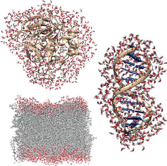
And thus regulating water position (the relative thermodynamics of hydrogen bonds) becomes an attractive proposition in the regulation of protein aggregation in dementia.
Lower water directly around proteins, increase viscosity and thus condensates facilitate phase separation.
Lower water directly around proteins, increase viscosity and thus condensates facilitate phase separation.
What helps, well visible 670 nm red light reduces viscosity in mitochondria interfacial water to increase free water molecules and enhance ATP synthase ability to generate more ATP. 

ROS also increase viscosity and inhibits ATP production.
Melatonin lowers viscosity by scavenging hydroxyl radical and ROS.
mdpi.com/1422-0067/24/6…
Melatonin lowers viscosity by scavenging hydroxyl radical and ROS.
mdpi.com/1422-0067/24/6…
This is beginning to make a lot more sense.
https://twitter.com/drkohilathas/status/1635987971873595392?s=20
• • •
Missing some Tweet in this thread? You can try to
force a refresh

